Copyright
©The Author(s) 2017.
World J Orthop. Oct 18, 2017; 8(10): 747-753
Published online Oct 18, 2017. doi: 10.5312/wjo.v8.i10.747
Published online Oct 18, 2017. doi: 10.5312/wjo.v8.i10.747
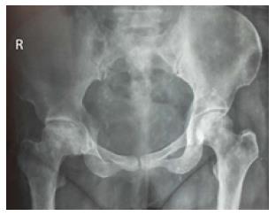
Figure 1 X-ray pelvis anteroposterior view shows collapsed right femoral head with sclerosis and subchondral lucencies, suggestive of osteonecrosis of right femoral head.
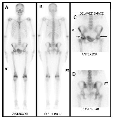
Figure 2 A 30-year-old female treated with chemotherapy for breast cancer was referred for 99mTc-MDP bone scan to evaluate cause of disabling hip pain.
Whole body (A and B) and delayed images (C and D) demonstrate photopenic areas in bilateral femoral heads (arrows) with increased osteoblastic activity surrounding the photopenic region in the right femoral head, suggestive of bilateral avascular necrosis. Increased osteoblastic activity in bilateral distal femora is likely due to biomechanical stress reaction due to altered gait.
Figure 3 Coronal single photon emission computed tomography (A), coronal computed tomography (B) and coronal fused single photon emission computed tomography/computed tomography images (C) of the patient mentioned in Figure 2 localizes the photopenic defects to head of bilateral femora.
The lucent areas with surrounding sclerosis in both femoral heads on low dose computed tomography (CT) component of single photon emission computed tomography/CT (B) increase the diagnostic confidence and specificity.
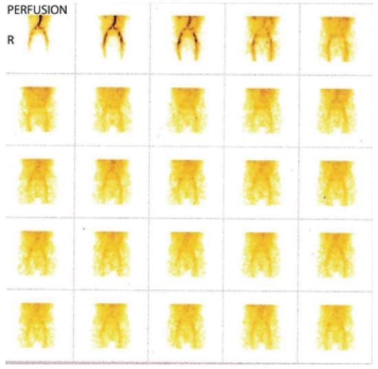
Figure 4 Perfusion phase of three-phase 99mTc-MDP bone scan of a patient with right side hip pain shows symmetrical flow of tracer in bilateral hips.
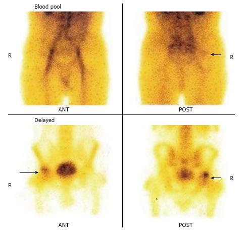
Figure 5 Blood pool images of the patient (as mentioned Figure 4) show minimally increased tracer uptake in the region of right hip.
The delayed images show increased tracer uptake in the right hip region with no definite photopenic area.
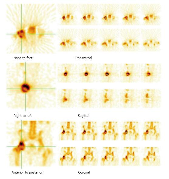
Figure 6 The single photon emission computed tomography images of the patient (as mentioned in Figure 4) show definite central photopenia with surrounding increased tracer uptake in the right femoral head (not evident on planar delayed images), suggestive of osteonecrosis of the right femoral head.
- Citation: Agrawal K, Tripathy SK, Sen RK, Santhosh S, Bhattacharya A. Nuclear medicine imaging in osteonecrosis of hip: Old and current concepts. World J Orthop 2017; 8(10): 747-753
- URL: https://www.wjgnet.com/2218-5836/full/v8/i10/747.htm
- DOI: https://dx.doi.org/10.5312/wjo.v8.i10.747


