Copyright
©The Author(s) 2017.
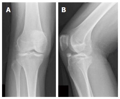
Figure 1 Initial radiographs of the right knee in the anteroposterior (A) and lateral (B) views.
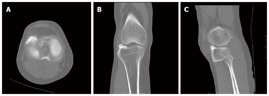
Figure 2 Initial computed tomography images of the right knee in the axial (A), coronal (B), and sagittal (C) planes.
Figure 3 Computed tomography images of the right knee after lesion recurrence in the axial (A), coronal (B), and sagittal (C) planes.
- Citation: Khalsa AS, Kumar NS, Chin MA, Lackman RD. Novel case of Trevor’s disease: Adult onset and later recurrence. World J Orthop 2017; 8(1): 77-81
- URL: https://www.wjgnet.com/2218-5836/full/v8/i1/77.htm
- DOI: https://dx.doi.org/10.5312/wjo.v8.i1.77


