Copyright
©The Author(s) 2016.
World J Orthop. Aug 18, 2016; 7(8): 494-500
Published online Aug 18, 2016. doi: 10.5312/wjo.v7.i8.494
Published online Aug 18, 2016. doi: 10.5312/wjo.v7.i8.494
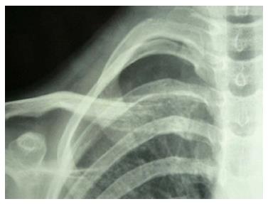
Figure 1 Radiograph of a patient with condensing osteitis showing expansion of the medial end of right clavicle and remodeling of bone.
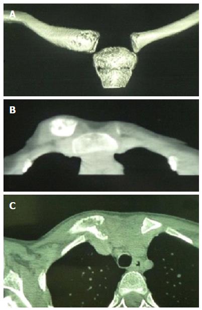
Figure 2 Computed tomography scan with three-dimensional reconstruction (A) of the right clavicle in a patient with condensing osteitis (B, C).
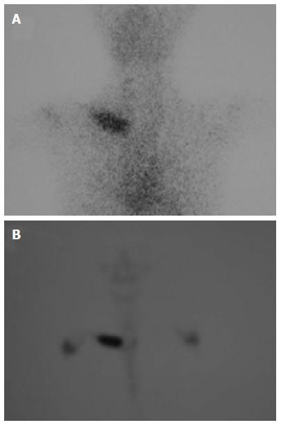
Figure 3 Triphasic bone scan showing strong, localized and isolated hyperfixation of the tracer at the level of the medial end of the right clavicle (A, B).
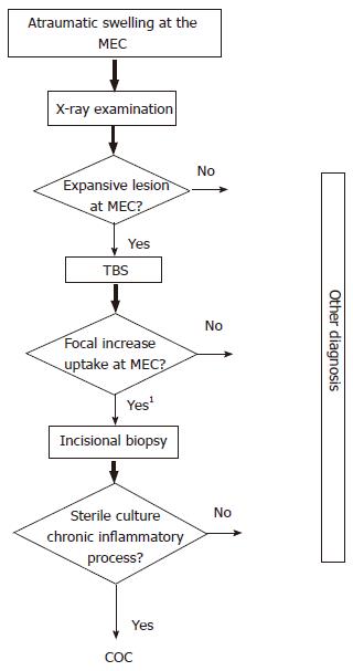
Figure 4 Diagnostic algorithm.
1If any doubt, fisrt perform MRI or CT scan. MRI: Magnetic resonance imaging; TBS: Triphasic bone scan; MEC: Medial end of calvicle; COC: Condensing osteitis of the clavicle.
- Citation: Andreacchio A, Marengo L, Canavese F. Condensing osteitis of the clavicle in children. World J Orthop 2016; 7(8): 494-500
- URL: https://www.wjgnet.com/2218-5836/full/v7/i8/494.htm
- DOI: https://dx.doi.org/10.5312/wjo.v7.i8.494


