Copyright
©The Author(s) 2016.
World J Orthop. Jul 18, 2016; 7(7): 452-457
Published online Jul 18, 2016. doi: 10.5312/wjo.v7.i7.452
Published online Jul 18, 2016. doi: 10.5312/wjo.v7.i7.452
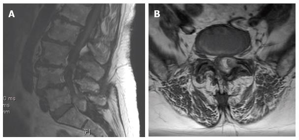
Figure 1 T2 weighted midsagittal (A) and axial (B) magnetic resonance imaging scan showed intraspinal extradural well delineated capsule containing a mixed signal intensity lesion on the left side at L5-S1 causing server canal stenosis with displacement of the thecal sac to the right side.
Figure 2 T1 weighted midsagittal (A) and axial (B) magnetic resonance imaging scan showed increased signal intensity with layering areas suggesting different stages of hematoma formation.
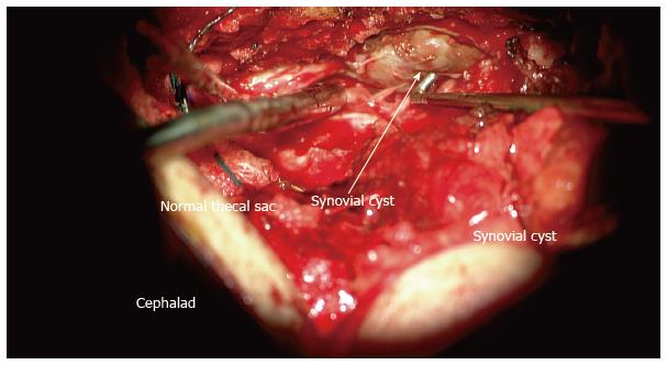
Figure 3 Intraoperative photograph taken by the surgical microscope showed a well-demarked brown color cyst that was adherent to the thecal sac (arrow), a hematoma was found inside the cyst.
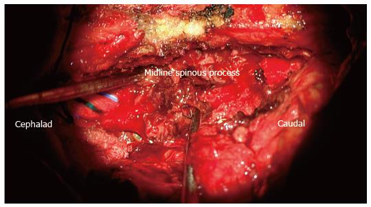
Figure 4 Intraoperative photograph taken by the surgical microscope showed hematoma inside the cyst (arrow).
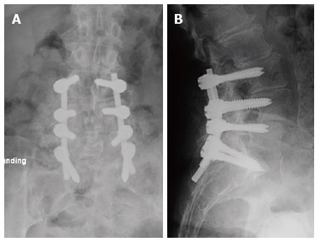
Figure 5 Postoperative anteroposterior and lateral roentgenograms showed L3-S1 instrumentation and posterolateral fusion.
- Citation: Elgafy H, Peters N, Lea JE, Wetzel RM. Hemorrhagic lumbar synovial facet cyst secondary to transforaminal epidural injection: A case report and review of the literature. World J Orthop 2016; 7(7): 452-457
- URL: https://www.wjgnet.com/2218-5836/full/v7/i7/452.htm
- DOI: https://dx.doi.org/10.5312/wjo.v7.i7.452


