Copyright
©The Author(s) 2016.
World J Orthop. Jun 18, 2016; 7(6): 401-405
Published online Jun 18, 2016. doi: 10.5312/wjo.v7.i6.401
Published online Jun 18, 2016. doi: 10.5312/wjo.v7.i6.401
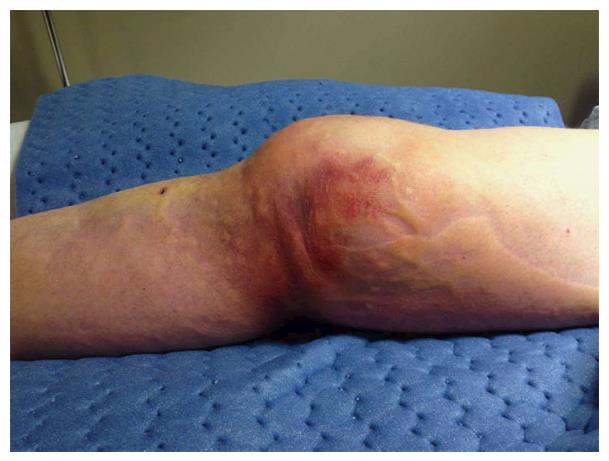
Figure 1 Clinical photograph showing the “pucker” sign with the knee in extension.
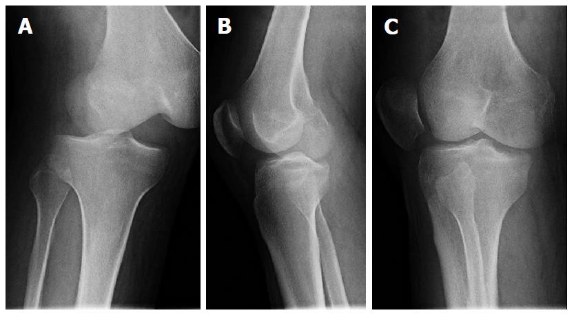
Figure 2 Supine radiographs showing posterolateral dislocation of the knee.
A: AP; B: Lateral; C: Oblique. Note similar projections (AP or lateral) of both the tibia and femur are not seen in any single view. AP: Anteroposterior.
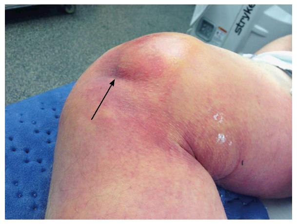
Figure 3 Clinical photograph under anesthesia showing accentuation of the “pucker” sign (black arrow) with flexion of the knee.
- Citation: Woon CY, Hutchinson MR. Posterolateral dislocation of the knee: Recognizing an uncommon entity. World J Orthop 2016; 7(6): 401-405
- URL: https://www.wjgnet.com/2218-5836/full/v7/i6/401.htm
- DOI: https://dx.doi.org/10.5312/wjo.v7.i6.401


