Copyright
©The Author(s) 2016.
World J Orthop. May 18, 2016; 7(5): 280-286
Published online May 18, 2016. doi: 10.5312/wjo.v7.i5.280
Published online May 18, 2016. doi: 10.5312/wjo.v7.i5.280
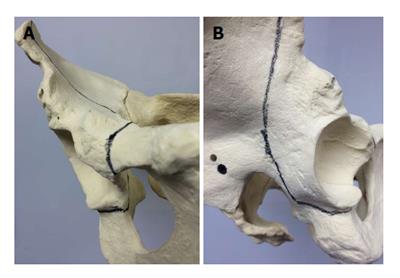
Figure 1 Location of osteotomy planes in the bernese periacetabular osteotomy.
Frontal (A) and lateral (B) views of the pelvis demonstrating placement of juxta-acetabular osteotome cuts (dark lines).
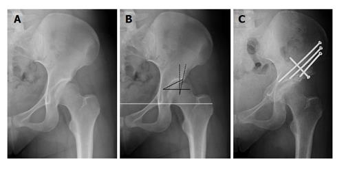
Figure 2 Acetabular radiographic measurements and correction.
Pre-operative (A) antero-posterior radiograph of a hip with classic dysplastic. Basic radiographic measurements of dysplasia (B) include the lateral center edge angle (dashed black line; measure of lateral acetabular coverage), Tönnis angle (solid black line; measure of sourcil angle); the solid white line is the inter-teardrop line, which is the reference for pelvic tilt in the coronal plane; an anterior center edge angle on a false profile view completes the basic radiographic work-up. The post-operative (C) antero-posterior radiographic view of the same hip demonstrating satisfactory acetabular reorientation to correct bony dysplasia.
- Citation: Kamath AF. Bernese periacetabular osteotomy for hip dysplasia: Surgical technique and indications. World J Orthop 2016; 7(5): 280-286
- URL: https://www.wjgnet.com/2218-5836/full/v7/i5/280.htm
- DOI: https://dx.doi.org/10.5312/wjo.v7.i5.280


