Copyright
©The Author(s) 2016.
World J Orthop. Mar 18, 2016; 7(3): 149-155
Published online Mar 18, 2016. doi: 10.5312/wjo.v7.i3.149
Published online Mar 18, 2016. doi: 10.5312/wjo.v7.i3.149
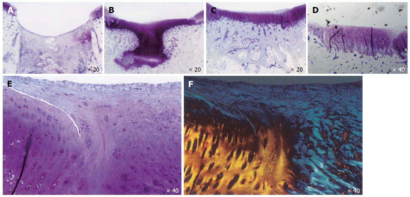
Figure 1 Articular cartilage healing in a microfracture model in adult rabbits.
Articular cartilage healing at day 7 (A), 21 (B), 42 (C), and day 84 (D-F). E and F: Lack of healing of reparative cartilage to “normal cartilage” is shown by toluidine blue and polarized light micrographs at day 84.
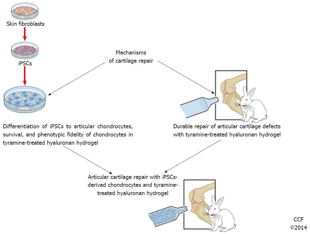
Figure 2 A broad outline for the use of induced pluripotent stem cells in articular cartilage repair.
iPSCs: Induced pluripotent stem cells.
- Citation: Lietman SA. Induced pluripotent stem cells in cartilage repair. World J Orthop 2016; 7(3): 149-155
- URL: https://www.wjgnet.com/2218-5836/full/v7/i3/149.htm
- DOI: https://dx.doi.org/10.5312/wjo.v7.i3.149


