Copyright
©The Author(s) 2016.
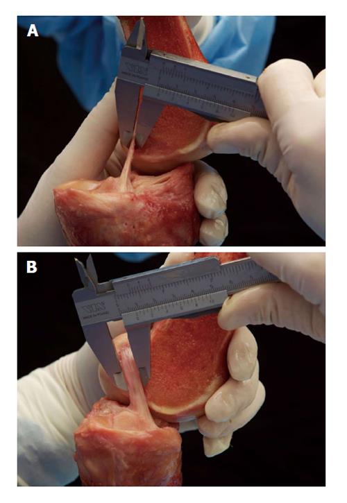
Figure 1 Śmigielski et al[17] measuring the thickness (A) and width (B) of the “ribbon-like” midsubstance of the anterior cruciate ligament.
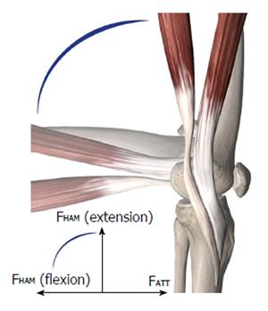
Figure 2 Semitendinosus and gracilis muscles with their insertions.
At 90° of flexion, the forces of the hamstrings (FHAM) are opposed to the anterior tibial translation (ATT) forces (FATT).
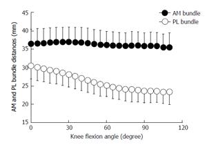
Figure 3 Distance between the femoral and tibial attachments of the anteromedial and posterolateral bundles of the anterior cruciate ligament during knee flexion (mean/SD) from Kondo et al[14].
AM: Anteromedial; PL: Posterolateral.
Figure 4 Shift of the center of the rotatory axis from the center/eminentia in an anterior cruciate ligament-intact knee (A) to a medial position (B).
Modified from Amis et al[24].
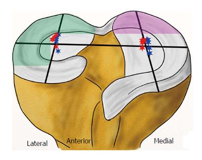
Figure 5 Lateral shift of femorotibial contact points at various flexion angles with intact anterior cruciate ligament (blue stars) and deficient anterior cruciate ligament (red stars) - modified from Li et al[40].
Increased contact stress after an anterior cruciate ligament rupture in the posterior lateral compartment during pivot shifting (green area) and in the posterior medial compartment during Lachman testing (violet area) - modified from Imhauser et al[41].
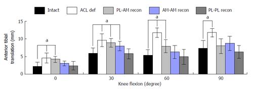
Figure 6 Anterior tibial translation under a simulated pivot shift test (combined 10 Nm valgus and 4 Nm internal rotational load, mean/SD, aP < 0.
05) as influenced by anterior cruciate ligament mismatch reconstruction of posterolateral and anteromedial bundles in a human cadaveric study by Herbort et al[8]. AM: Anteromedial; PL: Posterolateral; ACL: Anterior cruciate ligament.
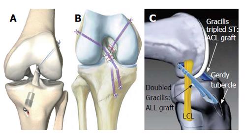
Figure 7 Novel anterior cruciate ligament repair techniques.
A: Anterior cruciate ligament (ACL) suture and dynamic augmentation (Ligamys™, copyright Mathys Ltd., Bettlach, Switzerland); B: Ligament bracing (from Heitmann et al[104]); C: ACL reconstruction with anterolateral ligament (ALL) augmentation (from Sonnery-Cottet et al[59]). ST: Semitendinosus, LCL: Lateral collateral ligament.
- Citation: Domnick C, Raschke MJ, Herbort M. Biomechanics of the anterior cruciate ligament: Physiology, rupture and reconstruction techniques. World J Orthop 2016; 7(2): 82-93
- URL: https://www.wjgnet.com/2218-5836/full/v7/i2/82.htm
- DOI: https://dx.doi.org/10.5312/wjo.v7.i2.82


