Copyright
©The Author(s) 2016.
World J Orthop. Dec 18, 2016; 7(12): 839-842
Published online Dec 18, 2016. doi: 10.5312/wjo.v7.i12.839
Published online Dec 18, 2016. doi: 10.5312/wjo.v7.i12.839
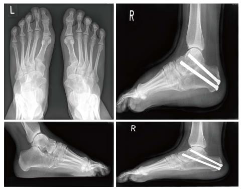
Figure 1 Radiographs of patient 1 demonstrating multiple tarsal coalitions and right foot radiographs following subtalar joint fusion.
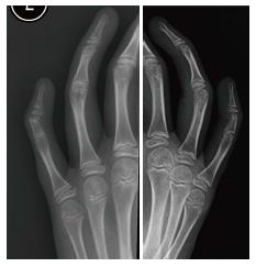
Figure 2 Radiographs of patient 2 (left) and patient 4 (right) demonstrating little fingers symphalangism of proximal a middle phalanx.
Patients are unable to flex the proximal interphalangeal joint of their little finger as opposed to the other fingers as demonstrated.
- Citation: Leonidou A, Irving M, Holden S, Katchburian M. Recurrent missense mutation of GDF5 (p.R438L) causes proximal symphalangism in a British family. World J Orthop 2016; 7(12): 839-842
- URL: https://www.wjgnet.com/2218-5836/full/v7/i12/839.htm
- DOI: https://dx.doi.org/10.5312/wjo.v7.i12.839


