Copyright
©The Author(s) 2016.
World J Orthop. Dec 18, 2016; 7(12): 793-800
Published online Dec 18, 2016. doi: 10.5312/wjo.v7.i12.793
Published online Dec 18, 2016. doi: 10.5312/wjo.v7.i12.793
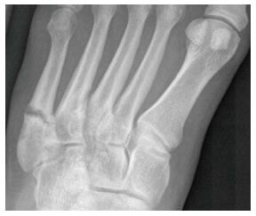
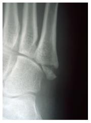
Figure 1 Radiograph of a Torg type II fifth metatarsal fracture.
Figure 2 Radiograph of a fifth metatarsal Torg type III fracture, which has nonunited.
Figure 3 Radiograph of a zone one fifth metatarsal fracture.
Figure 5 Radiograph showing healing in a nonunion treated with a percutaneous 3.
5-mm cortical lag screw.
- Citation: Bowes J, Buckley R. Fifth metatarsal fractures and current treatment. World J Orthop 2016; 7(12): 793-800
- URL: https://www.wjgnet.com/2218-5836/full/v7/i12/793.htm
- DOI: https://dx.doi.org/10.5312/wjo.v7.i12.793


