Copyright
©The Author(s) 2016.
World J Orthop. Dec 18, 2016; 7(12): 785-792
Published online Dec 18, 2016. doi: 10.5312/wjo.v7.i12.785
Published online Dec 18, 2016. doi: 10.5312/wjo.v7.i12.785
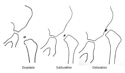
Figure 1 Drawings of the three types of the disease in infants.
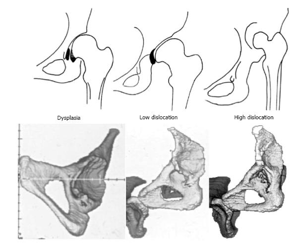
Figure 2 Drawings and three dimensional-computed tomography images of the 3 types of congenital hip disease in adults.
The black-colored areas in dysplasia represents the large osteophyte that covers the acetabular fossa and the medial marginal osteophyte of the femoral head (capital drop), and in low dislocation it the inferior part of the false acetabulum that is an osteophyte which begins at the level of the superior rim of the true acetabulum.
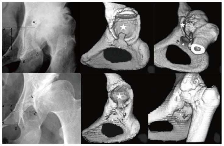
Figure 3 Images illustrating the two subtypes of low dislocation: B1 and B2.
Three points must be recognised on radiographs: (A) the superior limit of the true acetabulum; (B) the inferior point of the teardrop; (C) the most inferior point of the false acetabulum. Three dimensional-computed tomography scans may help to determine the superior limit of the true acetabulum, when it is not clear in plain radiographs. Asterisks depict false acetabulum and arrowheads true acetabulum.
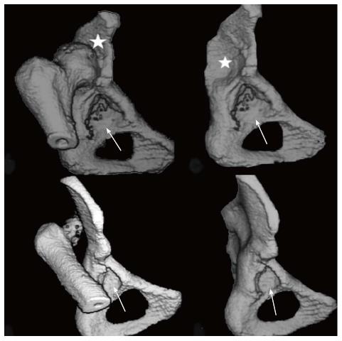
Figure 4 Three dimensional-computed tomography scans of the two subtypes of high dislocation.
Arrows indicate the true and asterisks the false acetabulum.
Figure 5 Development of dysplastic hips.
A: At the age of 3 years, when the child was first seen by her physician. An abduction frame was applied for 6 mo; B: At the age of 35 years, the patient had the first symptoms, pain and limping, due to the development of secondary hip osteoarthritis.
Figure 6 Radiographs and diagrams of a female patient with subluxation of the left hip in infancy, subsequently developed to low dislocation.
A: At the age of 2 years; B: Image, when the patient was 37 years old.
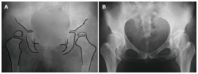
Figure 7 Images illustrating the case of a female patient with bilateral high dislocation.
A: Radiograph at the age of 2 years; B: Radiograph, at the age of 33 years, when the patient was consulted for bilateral total hip replacements.
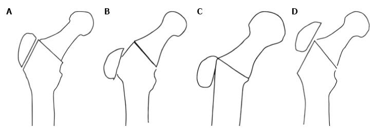
Figure 8 Drawings depicting.
A: Reattachment of the trochanter at its original bed; B: Distal reattachment of the trochanter, retaining contact with the distal part of the original bed; C: Reattachment of the trochanter on the lateral femoral cortex; D: Proximal reattachment of the trochanter.
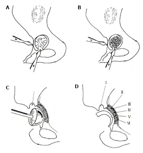
Figure 9 Cotyloplasty technique.
A: Comminuted fracture of the entire medial wall; B: Large amount of cancellous morselised graft placed between the fragments of the acetabular floor, onto the periosteum; C: Grafts moulded with a hemispherical pusher; D): Final appearance: I: Anchorage hole; II: Internal layer of the periosteum; III: Autogenous morselised graft; IV: Fragments of the acetabular floor; V: Cement mantle; VI: Offset-bore acetabular component.
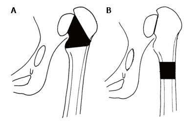
Figure 10 Drawings of the two alternatives of shortening of the femur.
A: At the level of femoral neck; B: Distal shortening at the level of femoral shaft.
- Citation: Hartofilakidis G, Lampropoulou-Adamidou K. Lessons learned from study of congenital hip disease in adults. World J Orthop 2016; 7(12): 785-792
- URL: https://www.wjgnet.com/2218-5836/full/v7/i12/785.htm
- DOI: https://dx.doi.org/10.5312/wjo.v7.i12.785


