Copyright
©The Author(s) 2016.
World J Orthop. Dec 18, 2016; 7(12): 776-784
Published online Dec 18, 2016. doi: 10.5312/wjo.v7.i12.776
Published online Dec 18, 2016. doi: 10.5312/wjo.v7.i12.776
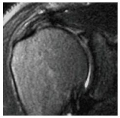
Figure 1 Magnetic resonance image of Bennett lesion and corresponding arthroscopic picture viewing posteriorly from anterosuperior portal.
Figure 2 Magnetic resonance image of a Type 2 SLAP tear with concomitant partial thickness rotator cuff tear.
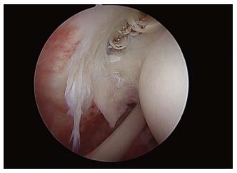
Figure 3 Partial thickness articular-sided tear of infraspinatus as viewed from posterior portal.
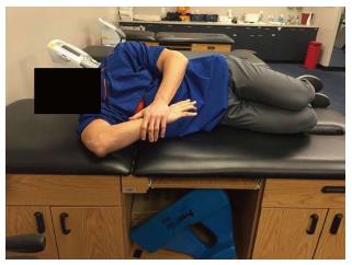
Figure 4 Demonstration of the “sleeper stretch”.
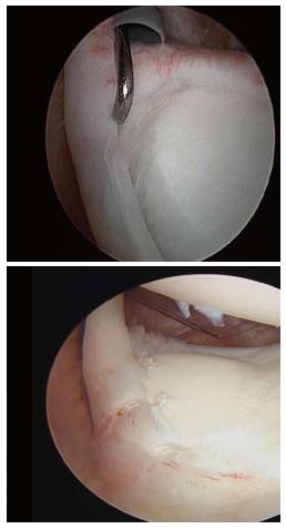
Figure 5 Type 2B SLAP tear s/p repair.
- Citation: Corpus KT, Camp CL, Dines DM, Altchek DW, Dines JS. Evaluation and treatment of internal impingement of the shoulder in overhead athletes. World J Orthop 2016; 7(12): 776-784
- URL: https://www.wjgnet.com/2218-5836/full/v7/i12/776.htm
- DOI: https://dx.doi.org/10.5312/wjo.v7.i12.776


