Copyright
©The Author(s) 2016.
World J Orthop. Nov 18, 2016; 7(11): 731-737
Published online Nov 18, 2016. doi: 10.5312/wjo.v7.i11.731
Published online Nov 18, 2016. doi: 10.5312/wjo.v7.i11.731
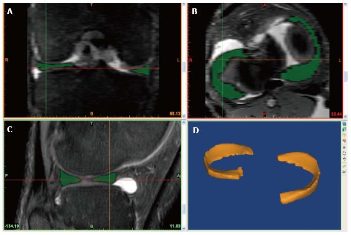
Figure 1 The user interface of the Materialise Interactive Medical Control System segmentation software program depicting the coronal view (A), the axial view (B), the sagittal view (C) and the three-dimentional reconstruction view (D).
Note the poorer contrast and pixelated images in coronal and axial windows as compared the sagittal window.
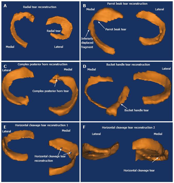
Figure 2 Three-dimensional reconstruction models showing an example of each configuration of meniscal tear identified in the study cohort.
Note the two illustrations provided for the horizontal cleavage tear. A: Radial tear reconstruction; B: Parrot bile tear reconstruction; C: Complex posterior horn reconstruction; D: Bucket handle tear reconstruction; E: Horizontal cleavage tear reconstruction 1; F: Horizontal cleavage tear reconstruction 2.
- Citation: Kruger N, McNally E, Al-Ali S, Rout R, Rees JL, Price AJ. Three-dimensional reconstructed magnetic resonance scans: Accuracy in identifying and defining knee meniscal tears. World J Orthop 2016; 7(11): 731-737
- URL: https://www.wjgnet.com/2218-5836/full/v7/i11/731.htm
- DOI: https://dx.doi.org/10.5312/wjo.v7.i11.731


