Copyright
©The Author(s) 2016.
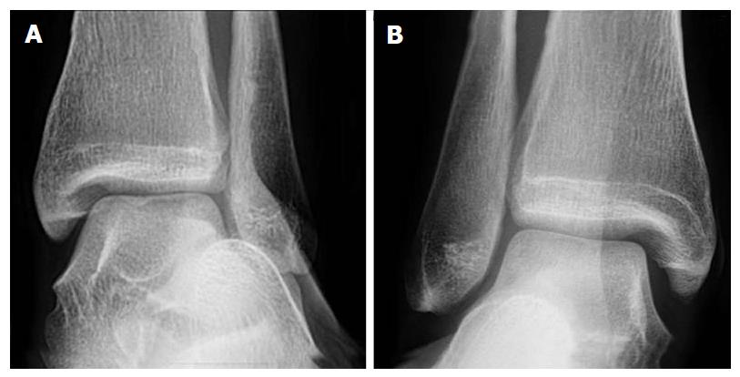
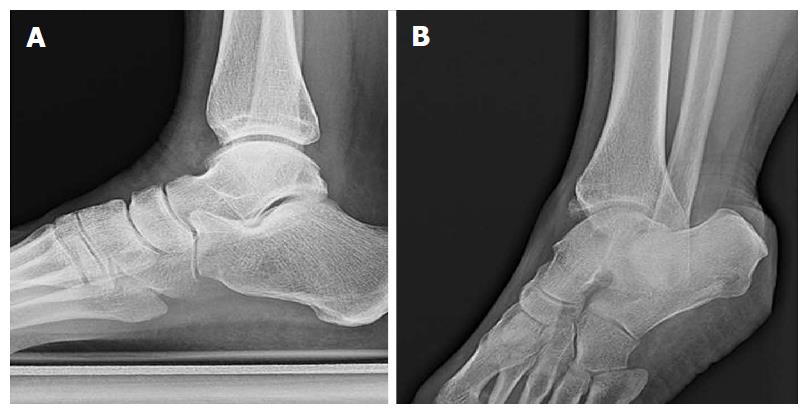
Figure 1 Plain radiographs of the left (A) and right (B) ankles of a single patient in the coronal plane.
Both ankles are under eversion stress. The right ankle was symptomatic. Only subtle syndesmotic gapping and widening of the medial clear space can be appreciated on the right compared to the left ankle.
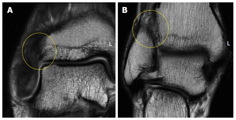
Figure 2 Coronal fast-spin echo proton density magnetic resonance images of the right ankle seen in Figure 1.
Disruption and remodeling of the anterior inferior tibiofibular ligament (A; yellow circle) and interosseous ligament (B; yellow circle) can be appreciated.
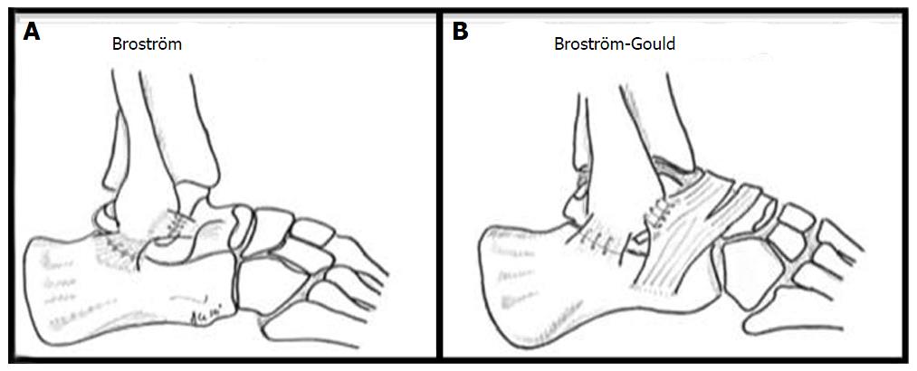
Figure 4 Illustrations of the Brostrom (A) and Modified Brostrom-Gould (B) surgical technique for lateral ligament reconstruction of the ankle.
Reproduced, with permission, from Prisk et al[108].
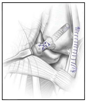
Figure 5 Illustration of the hybrid anatomic lateral ligament reconstruction[27].
A tendon autograft taken from the peroneus longus has been docked in the talus and distal fibula and remaining anterior talofibular ligament fibers have been sutured over the reconstruction, theoretically allowing proprioceptive fibers to aid in regaining functional stability. Illustration copyright of and reproduced with permission from Kennedy JG, MD. Reproduction without express written consent is prohibited. Reproduced, with permission, from Kennedy et al[40].
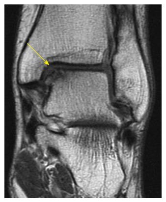
Figure 6 Coronal fast-spin echo proton density magnetic resonance image demonstrating an uncontained osteochondral lesion of the medial talar dome.
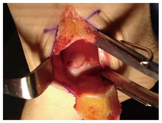
Figure 7 Intraoperative photograph of an autologous osteochondral graft transplanted into the medial talar dome.
Access was achieved via a medial malleolar osteotomy. The graft was gently tamped into position until flush with the surrounding surface.
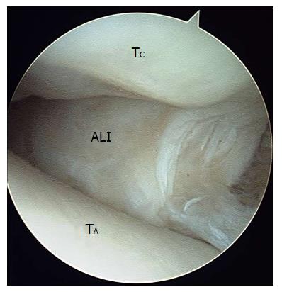
Figure 8 Arthroscopic image of soft tissue impingement the later gutter of the ankle joint.
ALI: Anterolateral soft tissue impingement including cicatraized tissue, entrapped lateral ligaments, and hypertrophied synovium; TA: Talar dome; TC: Tibial cartilage.
Figure 9 Standard lateral views about anterior tibial osteophytes.
Lateral plain radiograph of a right ankle with clinical suspicion of anteromedial impingement (A). An anteromedial radiographic view with 30 degrees of external rotation of the same ankle demonstrated an osteophyte on the anteromedial aspect of the distal tibia (B) that could not be appreciated on the lateral view.
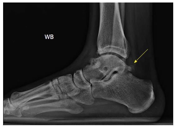
Figure 10 Lateral plain radiograph of a right ankle revealing an os trigonum, which was clinically symptomatic, as well as anterior tibial and talar osteophytes.
- Citation: Walls RJ, Ross KA, Fraser EJ, Hodgkins CW, Smyth NA, Egan CJ, Calder J, Kennedy JG. Football injuries of the ankle: A review of injury mechanisms, diagnosis and management. World J Orthop 2016; 7(1): 8-19
- URL: https://www.wjgnet.com/2218-5836/full/v7/i1/8.htm
- DOI: https://dx.doi.org/10.5312/wjo.v7.i1.8


