Copyright
©The Author(s) 2015.
World J Orthop. Oct 18, 2015; 6(9): 738-743
Published online Oct 18, 2015. doi: 10.5312/wjo.v6.i9.738
Published online Oct 18, 2015. doi: 10.5312/wjo.v6.i9.738
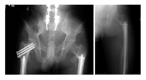
Figure 1 Anteroposterior and lateral radiographs of left femur showing peri-implant fracture involving the interlocking screw site in the subtrochanteric area.
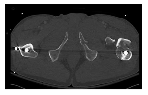
Figure 2 Axial computed tomography scan image showing the fracture at the screw insertion site.
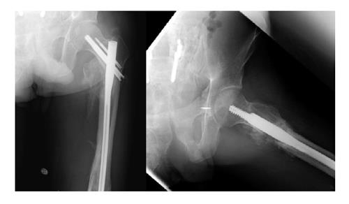
Figure 3 Anteroposterior and lateral radiographs six months after internal fixation of the peri-implant fracture.
Figure 4 Anteroposterior and lateral radiographs of right femur showing peri-implant fracture involving the Interlocking screw site in the subtrochanteric area.
Also seen is the femoral shaft fracture with interval healing.
Figure 5 Sagittal computed tomography scan image showing the fracture at the screw insertion site.
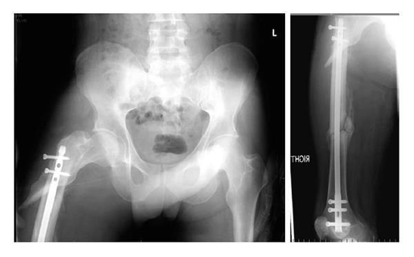
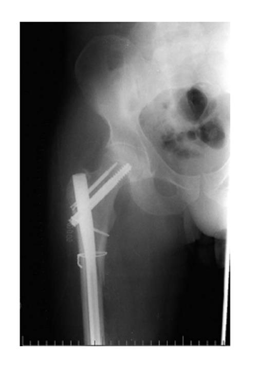
Figure 6 Anteroposterior radiograph of the pelvis post revision fixation.
Figure 7 Anteroposterior and lateral radiographs six months after internal fixation of the peri-implant fracture.
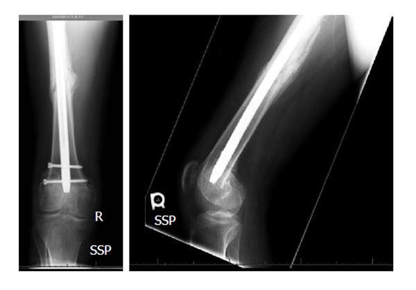
Figure 8 Anteroposterior and lateral radiographs showing healed diaphyseal femur fracture.
- Citation: Mounasamy V, Mallu S, Khanna V, Sambandam S. Subtrochanteric fractures after retrograde femoral nailing. World J Orthop 2015; 6(9): 738-743
- URL: https://www.wjgnet.com/2218-5836/full/v6/i9/738.htm
- DOI: https://dx.doi.org/10.5312/wjo.v6.i9.738


