Copyright
©The Author(s) 2015.
World J Orthop. Aug 18, 2015; 6(7): 559-563
Published online Aug 18, 2015. doi: 10.5312/wjo.v6.i7.559
Published online Aug 18, 2015. doi: 10.5312/wjo.v6.i7.559
Figure 1 Conventional radiograph at two months from onset revealing an irregular mass posterior to the distal humerus superior to the olecranon fossa.
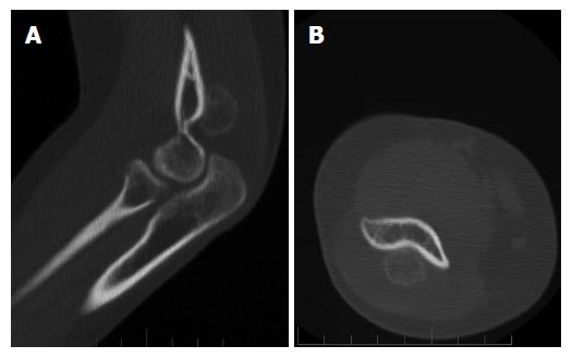
Figure 2 Sagittal (A) and axial (B) views demonstrating fine calcifications within the mass; no boney destruction was seen.
Figure 3 Computed tomography scan 11 wk after the initial imaging showing a 2.
0 cm × 1.9 cm osseous excrescence arising from the posterior distal humeral metaphysis.
Figure 4 Histologically the lesion was composed of woven bone in the background of mildly cellular bland spindle cells [haematoxylin and eosin, original magnification x 40 × (A), and × 100 (B)].
- Citation: Soni A, Weil A, Wei S, Jaffe KA, Siegal GP. Florid reactive periostitis ossificans of the humerus: Case report and differential diagnosis of periosteal lesions of long bones. World J Orthop 2015; 6(7): 559-563
- URL: https://www.wjgnet.com/2218-5836/full/v6/i7/559.htm
- DOI: https://dx.doi.org/10.5312/wjo.v6.i7.559


