Copyright
©The Author(s) 2015.
World J Orthop. Apr 18, 2015; 6(3): 322-330
Published online Apr 18, 2015. doi: 10.5312/wjo.v6.i3.322
Published online Apr 18, 2015. doi: 10.5312/wjo.v6.i3.322
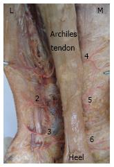
Figure 1 Two rows of perforators are distributed in the posterior lower leg sural region, originating from the peroneal artery (1, 2, and 3) in the posterolateral aspect and the posterior tibial artery (4, 5, and 6) in the posteromedial aspect, respectively.
Note the lateral peroneal artery perforators are developed more robust than that of the medial posterior tibial artery.
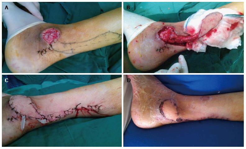
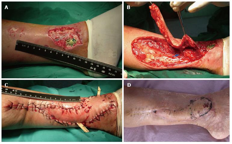
Figure 2 Distally based fasciocutaneous island flap for lateral malleolus coverage.
A: Pressure ulcer over the lateral malleolus in a 60-year-old male patient; B: Harvest of distally based fasciocutaneous island flap from the lateral sural region with a wide adipofascial pedicle; C: Flap inset to the recipient with tension-free; D: Final complete survival.
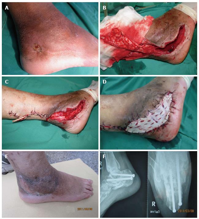
Figure 3 Distally based turnover adipofascial flap for calcaneal wound coverage.
A: Preoperative appearance of lateral calcaneal sinus; B: Distally based adipofascial flap harvest from the lateral sural region; C: The flap was turned 180-degrees over to fill the calcaneus cavity; D: Skin graft over the adipofascial flap; E: Appearance of postoperative 3 mo; F: Successful subtarlar joint fusion.
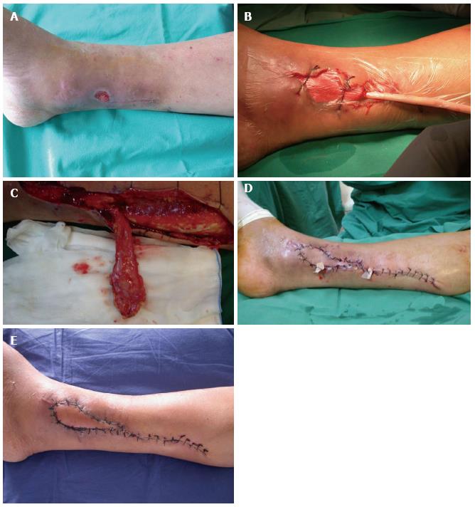
Figure 4 Perforator-plus fasciocutaneous flap for medial malleolar coverage.
A: Delayed wound infection after open pilon fracture; B: First stage management, debridement and vacuum-sealing-drainage; C: Perforator-plus fasciocutaneous flap harvest from the posteromedial sural region; D: Flap transfer and inset; E: Complete flap survival in two weeks.
Figure 5 Perforator pedicled propeller flap for pilon wound coverage.
A: Plate exposure in a 60-year-old woman with open pilon fracture; B: Posterior tibial artery perforator pedicled fasciocutaneous flap was raised, the perforator was skeletonized; C: The flap was propelled 180-degrees to the recipient site; D: Flap survival, with minor superficial marginal necrosis.
- Citation: Chang SM, Li XH, Gu YD. Distally based perforator sural flaps for foot and ankle reconstruction. World J Orthop 2015; 6(3): 322-330
- URL: https://www.wjgnet.com/2218-5836/full/v6/i3/322.htm
- DOI: https://dx.doi.org/10.5312/wjo.v6.i3.322


