Copyright
©The Author(s) 2015.
World J Orthop. Dec 18, 2015; 6(11): 850-863
Published online Dec 18, 2015. doi: 10.5312/wjo.v6.i11.850
Published online Dec 18, 2015. doi: 10.5312/wjo.v6.i11.850
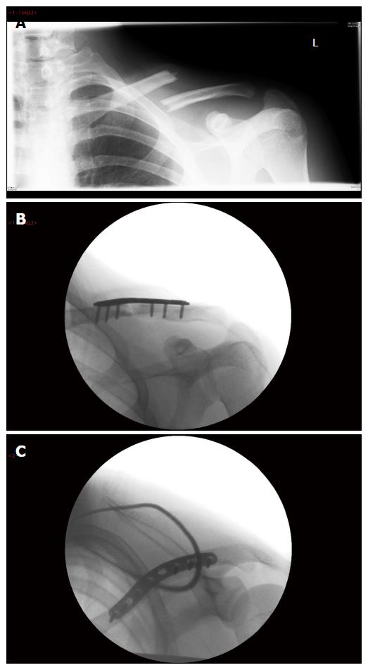
Figure 1 The management of a displaced mid diaphyseal clavicle fracture.
A: Pre-operative radiograph demonstrating complete displacement with shortening; B: Intra-operative antero-posterior radiograph; C: Intra-operative skyline radiograph.
Figure 2 The management of an undisplaced bimalleolar ankle fracture.
A, B: Pre-cast antero-posterior and lateral radiographs; C, D: Initial antero-posterior and lateral radiographs in cast; E, F: Two-week follow-up antero-posterior and lateral radiographs in cast.
Figure 3 The management of an undisplaced scaphoid waist fracture.
A, B: Pre-operative postero-anterior and 45-degree oblique radiographs; C: Intra-operative antero-posterior radiograph; D: Intra-operative lateral radiograph.
Figure 4 The management of a minimally displaced tibia shaft fracture.
A, B: Pre-cast antero-posterior and lateral radiographs; C, D: Initial antero-posterior and lateral radiographs in cast; E, F: Twelve-week post-operative antero-posterior and lateral radiographs.
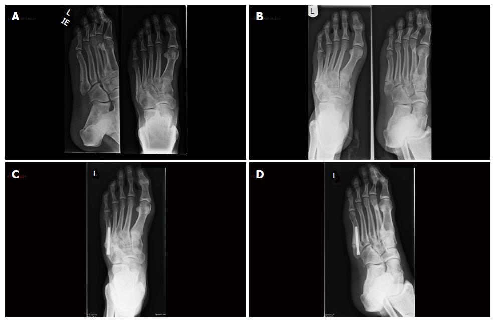
Figure 5 The management of an undisplaced fifth metatarsal jones fracture.
A: Initial Antero-posterior and oblique radiographs; B: Nine-week follow-up antero-posterior and oblique radiographs; C: Eight-week post-operative antero-posterior radiograph; D: Eight-week post-operative oblique radiograph.
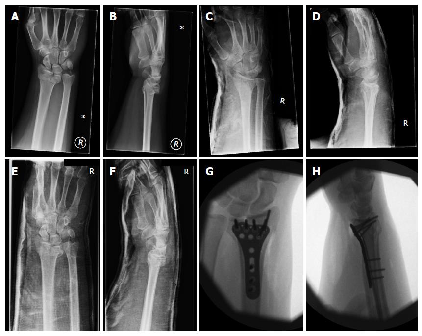
Figure 6 The management of the radiologically unstable distal radius fracture.
A, B: Initial postero-anterior and lateral radiographs radiographs; C, D: Post-manipulation postero-anterior and lateral radiographs; E, F: Two-week follow-up postero-anterior and lateral radiographs; G, H: Intra-operative antero-posterior and lateral radiographs.
- Citation: Robertson GA, Wood AM. Fractures in sport: Optimising their management and outcome. World J Orthop 2015; 6(11): 850-863
- URL: https://www.wjgnet.com/2218-5836/full/v6/i11/850.htm
- DOI: https://dx.doi.org/10.5312/wjo.v6.i11.850


