Copyright
©The Author(s) 2015.
World J Orthop. Nov 18, 2015; 6(10): 776-782
Published online Nov 18, 2015. doi: 10.5312/wjo.v6.i10.776
Published online Nov 18, 2015. doi: 10.5312/wjo.v6.i10.776
Figure 1 Anterior-posterior radiograph of the pelvis in a 24-year-old female with sickle cell anemia and long-standing, disabling bilateral hip pain.
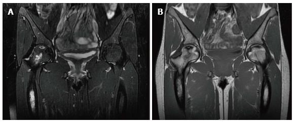
Figure 2 T2-weighted (A) and T1-weighted (B) coronal magnetic resonance imaging sequences demonstrating evidence of bilateral hip avascular necrosis and marrow changes consistent with sickle cell anemia.
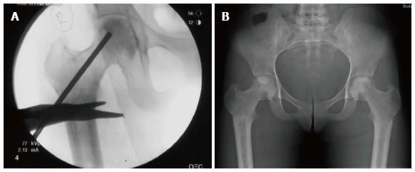
Figure 3 Intra-operative anterior-posterior fluoroscopic view (A) during a core decompression of the right hip, post-operative anterior-posterior radiograph of the pelvis (B) after bilateral core decompression.
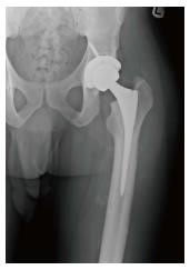
Figure 4 Anterior-posterior radiograph of the left hip in a 36-year-old male status post total hip arthroplasty for end-stage degenerative arthritis secondary to avascular necrosis.
- Citation: Kamath AF, McGraw MH, Israelite CL. Surgical management of osteonecrosis of the femoral head in patients with sickle cell disease. World J Orthop 2015; 6(10): 776-782
- URL: https://www.wjgnet.com/2218-5836/full/v6/i10/776.htm
- DOI: https://dx.doi.org/10.5312/wjo.v6.i10.776


