Copyright
©2014 Baishideng Publishing Group Inc.
World J Orthop. Sep 18, 2014; 5(4): 412-424
Published online Sep 18, 2014. doi: 10.5312/wjo.v5.i4.412
Published online Sep 18, 2014. doi: 10.5312/wjo.v5.i4.412
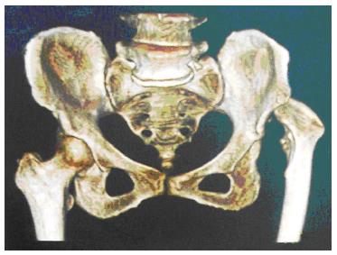
Figure 1 On the right side hip is normally developed and on the left side the acetabulum is underdeveloped, shallow and lacking bone stock medially and at the level of normal (ideal) acetabular roof.
The femoral head is more proximal (dislocated) with increased anteversion, shorter neck and narrower and straighter femoral canal.
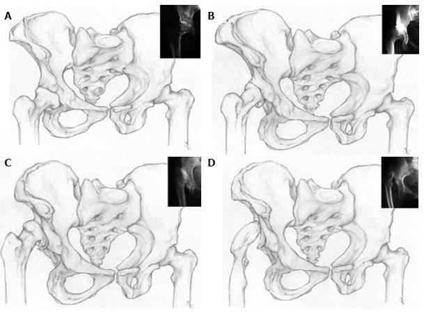
Figure 2 Left hip is normal, right hip is dysplastic.
A: Crowe type 1-proximal head subluxation is less than 50% of vertical diameter of the femoral head (less than 10% of the pelvic height); B: Crowe type 2-proximal head subluxation is between 50% and 75% of vertical diameter of the femoral head (between 10% and 15% of the pelvic height); C: Crowe type 3-proximal head subluxation is between 75% and 100% of vertical diameter of the femoral head (between 15% and 20% of the pelvic height); D: Crowe type 4-proximal head dislocation with proximal movement of the femoral head for more than 100% of vertical diameter of the femoral head (head is moved proximally for more than 20% of the pelvic height).
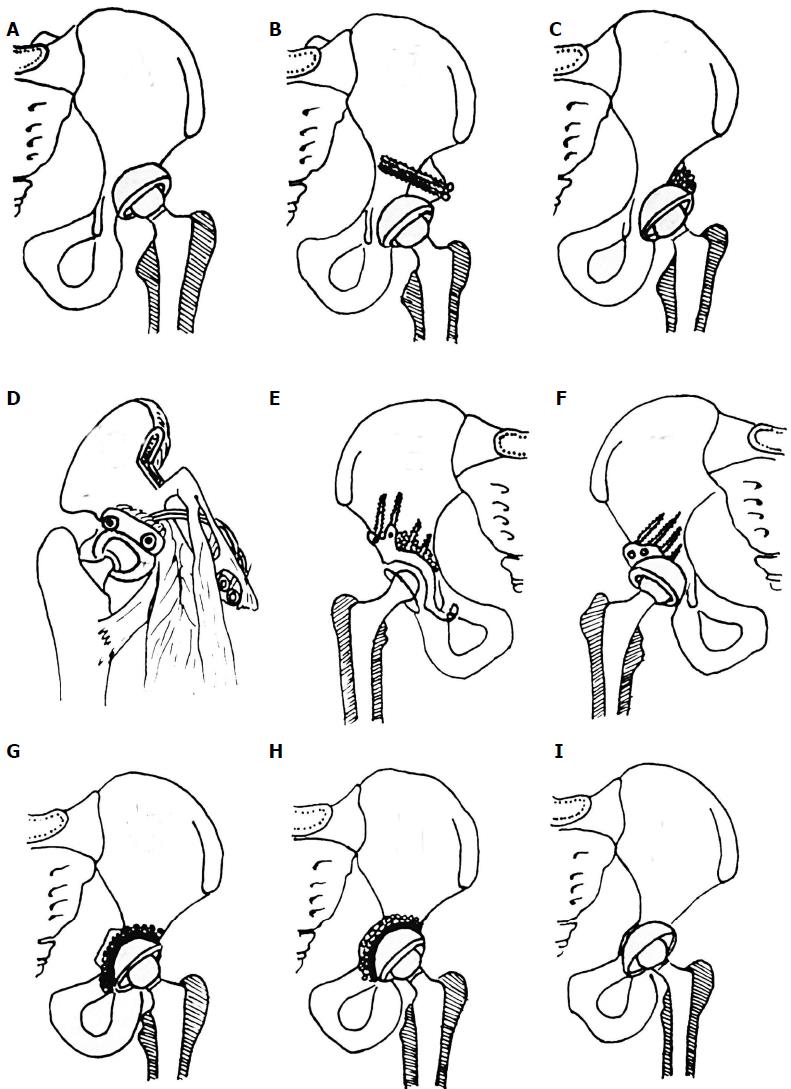
Figure 3 Different options for acetabular reconstruction.
A: Higher placement of the acetabular cup; B: Placement of the acetabular component in the anatomical position and augmentation of the superior segmental defect with structural autograft or allograft fixed with screws; C: Placement of the acetabular component in the anatomical position and spongioplasty of the acetabular roof for smaller uncovered areas (30%-40%); D: Anatomical position of acetabular cup and augmentation of the superior segmental defect with vascularised iliac graft; E: Reinforcement ring with the hook in combination with autologous graft augmentation for cases with severe bone-stock deficiencies. Anatomic hip centre is reconstructed by positioning the hook around the inferior margin of the acetabular floor. The hook prevents high or lateral placement of the ring and helps adequate coverage of the polyethylene liner, regardless of the degree of anatomical deformity; F: Acetabular bone stock deficiency can be managed with specially constructed acetabular components or using special 3-dimensional porous materials which simulates bone structure and allow faster and better endoprosthesis-bone integration. For that purpose trabecular metal (tantalum) is used in form of acetabular cup or trabecular metal augments. Oblong-shaped cementless implants can be used for acetabular reconstruction; G: Cotyloplasty with chisel - intentional medial wall fracture using osteotome with cup placement beyond the ilioischial line with bone grafting; H: Cotyloplasty with reamer - first, perforation of the medial acetabular wall with a reamer is performed, then acetabulum is filled with a large amount of autogenous cancelous bone graft and cup is cemented in position without pressure; I: Cotyloplasty without spongioplasty - implantation of porous-coated cementless acetabular components without spongioplasty.
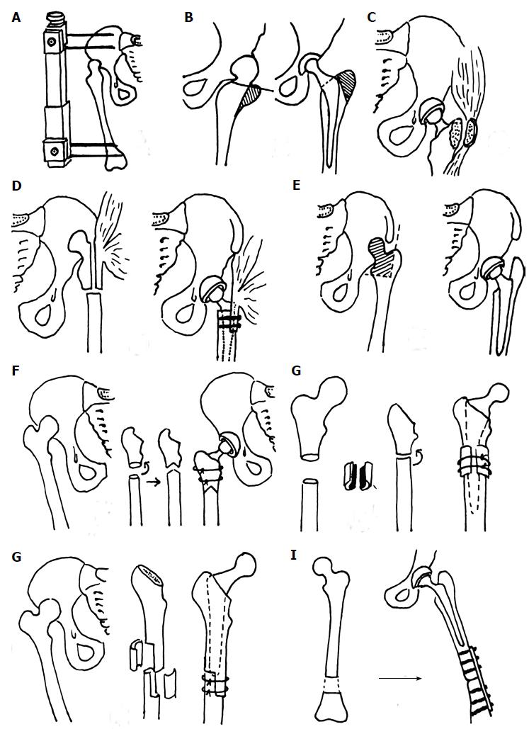
Figure 4 Different types of femoral reconstruction options.
A: Wagner’s apparatus for preoperative skeletal traction to reduce high congenital dislocation of the hip before total hip arthroplasty; B: Trochanteric osteotomy in total hip arthroplasty; C: Delimar et al[7] modification of the direct lateral approach to the hip. Anterior half of the continuous tendon is detached either by cautery or with a chisel. If the chisel is used, a thin layer of bone from the greater trochanter remains attached to the continuous tendon of the gluteus medius and the vastus lateralis. The posterior half of the continuous tendon of the gluteus medius and the vastus lateralis is always detached with the chisel leaving a bone flake of at least 2 to 3 mm thickness attached to tendons. In that way, the abductor muscles are stripped from the greater trochanter and there is no trochanteric osteotomy during the approach, which allows preservation of the continuity of the abductor muscles; D: Paavilainen's procedure of metaphyseal shortening osteotomy combined with distal sliding of the greater trochanter with intact attachment of the abductor muscles; E: Progressive femoral shortening at the level of lesser trochanter; greater trochanter remains intact, thus providing better functional results; F: Combined procedure of femoral subtrochanteric shortening with derotational double-chevron osteotomy. Transverse osteotomy was first performed, followed by rotational alignment in order to correct anteversion. Later, double chevron osteotomy was performed. Such method allows intraoperative derotation and shortening adjustment; G: Subtrochanteric osteotomy - modified technique; osteotomy sites were covered with onlay grafts of the excised fragments and fixed with two cerclage wires; H: Diaphyseal step-cut shortening osteotomy performed after reaming and stabilized with two to three cerclage bands with or without bone grafting. After stabile fixation, intramedullary reaming is done until optimal cortical contact is achieved, especially distal to the osteotomy site; I: Distal femur shortening procedure. First, total hip arthroplasty with acetabulum in anatomic position is performed followed by the femoral shortening that is done distal to stem so that the first screw of the plate would be more than 2 cm from the stem. Later, plate fixation of the femoral osteotomy site was performed.
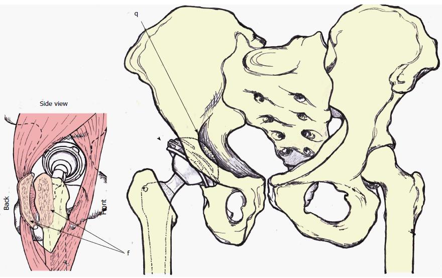
Figure 5 Anterior-posterior and latero-lateral (side) view of the author preferred method of treatment.
Anterior-posterior view - acetabular cup is medialized (cotyloplasty) so that the dome of the cup is protruding beyond Kohler’s line inside the pelvis (q marked with single arrow). Superolateral part of the cup is uncovered by the bone (marked with arrowhead). The cup is usually additionally secured with the screws (not show on the picture). latero-lateral (side) view-posterior part of the gluteus medius and vastus lateralis together with the external rotators are detached with the chisel on a thin flake of bone (f marked with double arrows). This is a modified direct lateral approach.
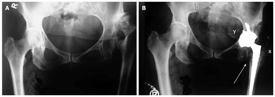
Figure 6 X-rays of patient with Crowe type 4 dysplasia on the left side and normal hip on right side.
A: Preoperative X-ray with secondary osteoarthritis due to dysplasia, neoacetabulum formed superolaterally from original, true acetabulum and significant leg length discrepancy; B: Postoperative X-ray with implanted uncemented acetabular cup and femoral stem. Acetabular cup is protruding beyond the Kohler’s line inside the pelvis (marked with y) and secured with 3 additional screws. Lesser trochanter is brought distally to the normal level so there is no leg length discrepancy postoperatively (marked with a single arrow). Modified direct lateral approach was used and posterior part of the gluteus medius and vastus lateralis together with the external rotators were detached with the chisel on a thin flake of bone, now they are completely attached and healed to greater trochanter (marked with x).
- Citation: Bicanic G, Barbaric K, Bohacek I, Aljinovic A, Delimar D. Current concept in dysplastic hip arthroplasty: Techniques for acetabular and femoral reconstruction. World J Orthop 2014; 5(4): 412-424
- URL: https://www.wjgnet.com/2218-5836/full/v5/i4/412.htm
- DOI: https://dx.doi.org/10.5312/wjo.v5.i4.412


