Copyright
©2014 Baishideng Publishing Group Inc.
World J Orthop. Jul 18, 2014; 5(3): 272-282
Published online Jul 18, 2014. doi: 10.5312/wjo.v5.i3.272
Published online Jul 18, 2014. doi: 10.5312/wjo.v5.i3.272
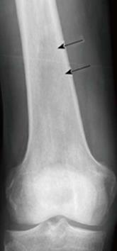
Figure 1 X-ray of an osseous myeloma lesion.
Conventional X-ray of the right femoral bone showing an osteolytic lesion (arrows) representing an osseous myeloma manifestation.
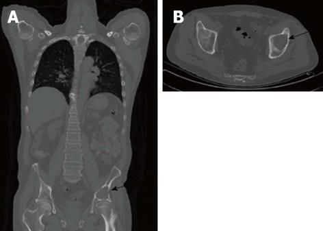
Figure 2 Computed tomography of an osseous myeloma lesion.
Computed tomography in coronal (A) and transversal views (B) showing an osteolytic lesion in the left iliac bone (arrows) representing an osseous myeloma manifestation.
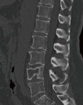
Figure 3 Computed tomography of an osseous myeloma lesion.
Computed tomography in sagittal view showing an osteolytic lesion in L4 with associated pathologic fracture.
Figure 4 Whole-body magnetic resonance imaging of an osseous myeloma lesion.
Whole-body magnetic resonance imaging: short-tau-inversion-recovery sequence (A), T1-weighted image (B) and T1-weighted image with fat suppression after contrast administration (C) showing an osseous lesion in L4 (arrows) representing an osseous myeloma manifestation.
Figure 5 Positron emission tomography/computed tomography of an osseous myeloma lesion.
Transversal computed tomography (CT) (A), 18F-fluorodeoxyglucose positron emission tomography (PET) (B) and fused PET/CT (C) showing an osteolytic lesion in the left iliac bone with cortical destruction representing an osseous myeloma manifestation.
Figure 6 Positron emission tomography/computed tomography of extraosseous myeloma lesions.
Transversal computed tomography (CT) (A), 18F-fluorodeoxyglucose positron emission tomography (PET) (B) and fused PET/CT (C) showing extraosseous myeloma manifestations (arrows).
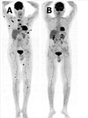
Figure 7 Positron emission tomography for therapy monitoring.
Whole-body maximum-intensity-projection positron emission tomography (PET) images before and after stem cell transplantation showing extensive osseous and extraosseous myeloma manifestations before therapy (A) and complete resolution on PET after therapy (B).
- Citation: Derlin T, Bannas P. Imaging of multiple myeloma: Current concepts. World J Orthop 2014; 5(3): 272-282
- URL: https://www.wjgnet.com/2218-5836/full/v5/i3/272.htm
- DOI: https://dx.doi.org/10.5312/wjo.v5.i3.272


