Copyright
©2014 Baishideng Publishing Group Co.
World J Orthop. Apr 18, 2014; 5(2): 146-150
Published online Apr 18, 2014. doi: 10.5312/wjo.v5.i2.146
Published online Apr 18, 2014. doi: 10.5312/wjo.v5.i2.146
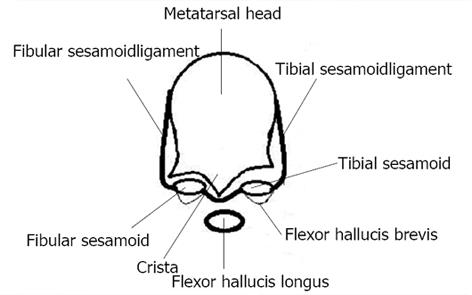
Figure 1 Diagrammatic representation of a cross section through the distal metatarsal and sesamoids.
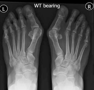
Figure 2 X-ray to demonstrate a case of sesamoid coalition.
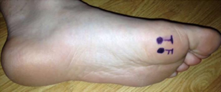
Figure 3 Photograph to show surface markings of the tibial (T) and fibular (F) sesamoids.
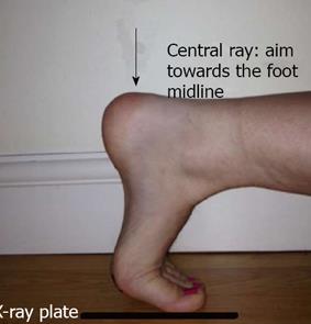
Figure 4 Photograph to show patient positioning to obtain a sesamoid axial view.
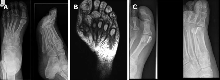
Figure 5 Symptomatic non-union may be treated with percutaneous screw fixation, open fixation or open bone grafting.
A: X-ray to show a symptomatic non-union of a sesamoid fracture; B: Magnetic resonance imaging scan demonstrating fracture of the sesamoid; C: Post-operative X-ray following fixation of a non-union sesamoid fracture.
- Citation: Sims AL, Kurup HV. Painful sesamoid of the great toe. World J Orthop 2014; 5(2): 146-150
- URL: https://www.wjgnet.com/2218-5836/full/v5/i2/146.htm
- DOI: https://dx.doi.org/10.5312/wjo.v5.i2.146


