Copyright
©2013 Baishideng Publishing Group Co.
World J Orthop. Oct 18, 2013; 4(4): 167-174
Published online Oct 18, 2013. doi: 10.5312/wjo.v4.i4.167
Published online Oct 18, 2013. doi: 10.5312/wjo.v4.i4.167
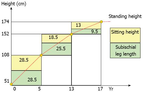
Figure 1 Sitting and standing height.
From birth to age five, the development of the trunk is substantial. Between age 5 and puberty, the lower extremities grow more than the trunk and the spinal growth rate decreases from 2.2 to 1.1 cm/year. During puberty, the trunk grows more than the lower extremities. At the beginning of puberty, the remaining standing height is approximately 20 cm, of which 2/3 is at the level of the trunk and 1/3 is at the level of the lower extremities. At Risser I (menarche), the remaining growth of the trunk is approximately 3 to 4 cm.
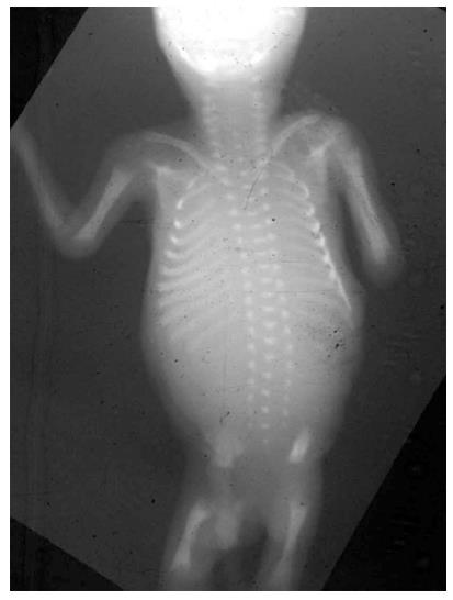
Figure 2 Spine at birth.
At birth, only 30% of the spine is ossified. At birth, the T1-S1 segment measures approximately 20 cm and reaches 45 cm at skeletal maturity.
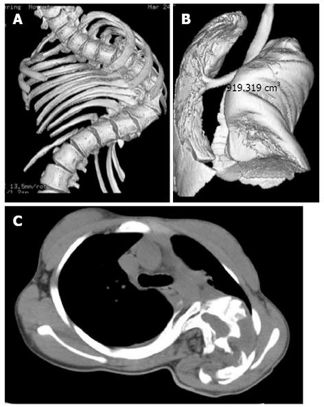
Figure 3 Thoracic cage distortion and lung compression.
Lung (A) and thoracic cage (B) growth volumes increase in a non-linear fashion over the first twenty years of life. The volumes of both structures are proportional to height in the absence of spinal disease. Spinal deformities alter normal spine and thorax growth, prevent the lungs from expanding and lead to thoracic insufficiency syndrome (C).
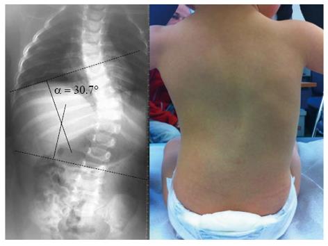
Figure 4 Early-onset spinal deformity.
Thirteen-month-old female with progressive infantile scoliosis.
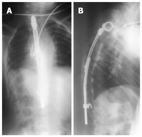
Figure 5 Controlled growth.
Eight-year-old male with congenital scoliosis treated with “spine to rib” vertical expandable prosthetic titanium rib. Treatment of the growing spine is a unique challenge and involves preservation of the thoracic spine, thoracic cage, and lung growth without reducing spinal motion.
- Citation: Canavese F, Dimeglio A. Normal and abnormal spine and thoracic cage development. World J Orthop 2013; 4(4): 167-174
- URL: https://www.wjgnet.com/2218-5836/full/v4/i4/167.htm
- DOI: https://dx.doi.org/10.5312/wjo.v4.i4.167


