Copyright
©2013 Baishideng Publishing Group Co.
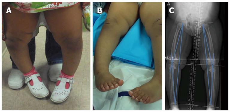
Figure 1 The girl had the diagnosis of bilateral infantile tibia vara with bilateral internal tibial torsion.
A: 3 years old girl, standing showing genu Varum; B: Pre operative clinical picture showing the marked internal rotation of both lower extremities. Note the relation between the feet direction and the patella (outlined by skin marker); C: Pre operative radiograph showing the gneu varum, mechanical axis deviation and the proximal medial tibial physeal changes.
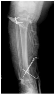
Figure 2 Immediate post operative radiograph for the right leg showing application of the lateral plate to proximal tibial growth plate and distal tibial/fibular osteotomy fixed by 3 K wires.
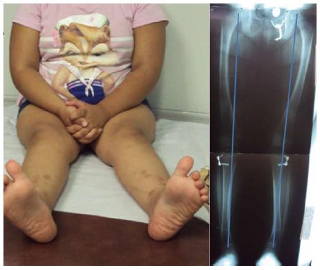
Figure 3 Ten months post operative showing.
A: Correction of both the genu varum and the internal rotation of the leg (clinical picture); B: Correction of the varus deformity and healing of the osteotomy sites (radiograph).
- Citation: Abdelgawad AA. Combined distal tibial rotational osteotomy and proximal growth plate modulation for treatment of infantile Blount’s disease. World J Orthop 2013; 4(2): 90-93
- URL: https://www.wjgnet.com/2218-5836/full/v4/i2/90.htm
- DOI: https://dx.doi.org/10.5312/wjo.v4.i2.90


