Copyright
©2013 Baishideng Publishing Group Co.
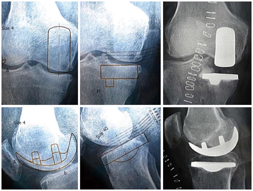
Figure 1 Example of a preoperative surgical plan of a medial unicompartmental knee arthroplasty right knee.
Weight-bearing radiographs are templated against acetate phantoms. Immediate post-operation radiographs show correct positioning of the prosthetic implants.
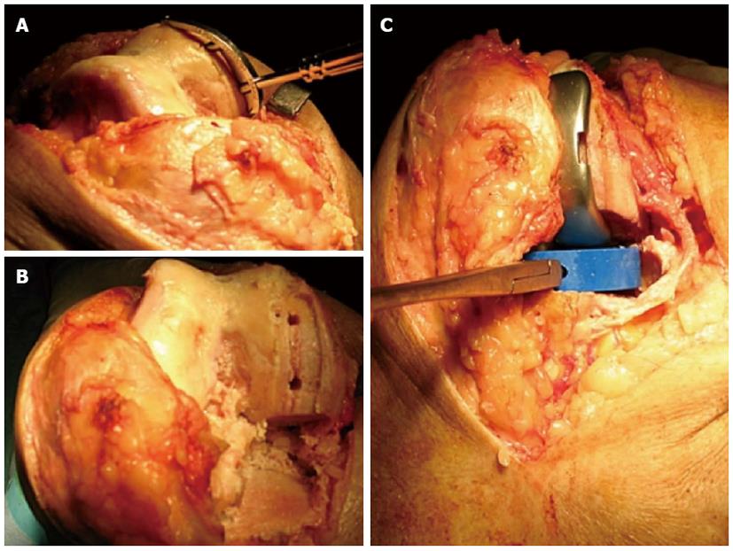
Figure 2 Knee bone cuts and positioning of trial components.
A: A curved instrument available in different sizes allows to check the curvature of the condylus to prosthetize along with the amount of bone to remove; B: Femoral and tibial bony cuts. At this stage of the operation is essential to check eventual meniscal fragments, bony particulate and bony prominences that is made possible through a standard parapatellar approach; C: Femoral and tibial trials inserted with patella in place. Accurate trials size to choose definitive implants must be carefully checked.
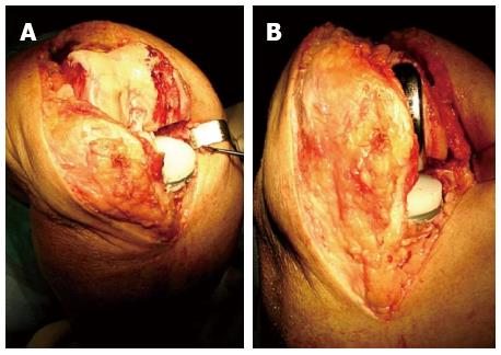
Figure 3 Cemented prosthetic components in place and patellar tracking assessment.
A: Cemented tibial metal-back component in place with proper thickness of polyethylene insert; B: Cemented femoral and tibial components inserted along with patella in place. At this moment it is possible to verify ligament balance and patellar tracking.
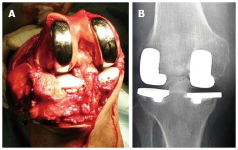
Figure 4 Bi-unicompartmental knee arthroplasty.
A: In selected cases, bi-unicompartmental knee replacement is a feasible prosthetic solution that allows to maintain ligamentous compartments; B: This permits to have a more physiologic knee functionality, replacing only the affected parts of the articulation.
- Citation: Salvi AE, Florschutz AV. Unicompartmental knee prosthetization: Which key-points to consider? World J Orthop 2013; 4(2): 58-61
- URL: https://www.wjgnet.com/2218-5836/full/v4/i2/58.htm
- DOI: https://dx.doi.org/10.5312/wjo.v4.i2.58


