Copyright
©2013 Baishideng Publishing Group Co.
Figure 1 Endplate disruption grading method schematic diagram.
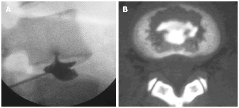
Figure 2 Discography and computed tomography.
A: Discography showing a radial disruption on the lower endplate of L4 vertebra and that the contrast medium flows into the cancellous bone of the lower endplate of L4 vertebra through the fissure; B: Computed tomography scan showing the contrast medium dispersed in the lower endplate of L4 vertebra, with Grade 4 endplate disruption.
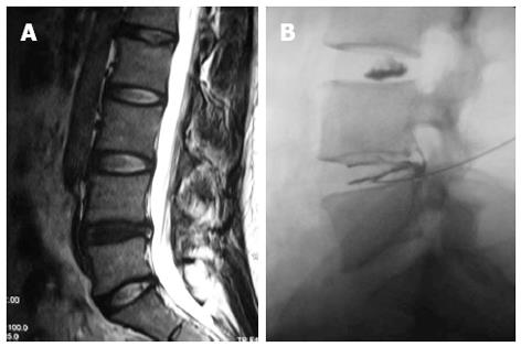
Figure 3 Magnetic resonance imaging and discography.
A: A 35-year-old woman had a 5-year history of low back pain. Sagittal T2 weighted magnetic resonance imaging showed L4/5 disc degeneration with a high intensity zone in the posterior annulus fibrosus; B: Discography showed L4/5 disc disruption with exact pain reproduction. After discography, 10 mg methylene blue was injected into the painful disc through discographic needle. Low back pain was almost totally relieved. No recurrence was observed at a 12-mo follow-up interval.
- Citation: Peng BG. Pathophysiology, diagnosis, and treatment of discogenic low back pain. World J Orthop 2013; 4(2): 42-52
- URL: https://www.wjgnet.com/2218-5836/full/v4/i2/42.htm
- DOI: https://dx.doi.org/10.5312/wjo.v4.i2.42


