Copyright
©2012 Baishideng Publishing Group Co.
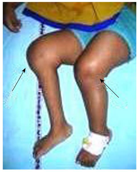
Figure 1 Clinical photo of patient showing swelling of bilateral knees (arrow).
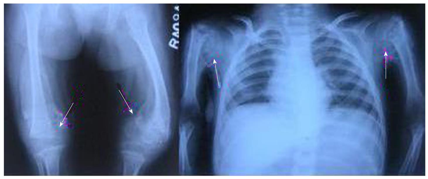
Figure 2 Plain radiograph of bilateral bilateral knees shoulder and showing epiphyseal separation (marked with arrow).
Figure 3 The follow-up radiograph shows normal alignment of epiphysis.
- Citation: Gupta S, Kanojia R, Jaiman A, Sabat D. Scurvy: An unusual presentation of cerebral palsy. World J Orthop 2012; 3(5): 58-61
- URL: https://www.wjgnet.com/2218-5836/full/v3/i5/58.htm
- DOI: https://dx.doi.org/10.5312/wjo.v3.i5.58


