Copyright
©2012 Baishideng Publishing Group Co.
World J Orthop. Dec 18, 2012; 3(12): 204-211
Published online Dec 18, 2012. doi: 10.5312/wjo.v3.i12.204
Published online Dec 18, 2012. doi: 10.5312/wjo.v3.i12.204
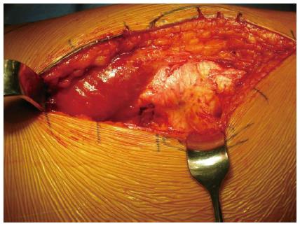
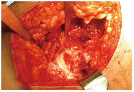
Figure 1 Straight lateral approach is shown for Ganz surgical dislocation.
It is extremely important to well visualize the posterior border of the greater trochanter prior to proceeding for the trochanteric flip osteotomy.
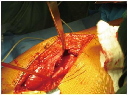
Figure 2 Trochanteric flip osteotomy begins at the posterior border of the greater trochanter and is carried anterior exiting superficial to the piriformis fossa and distal to the vastus ridge.
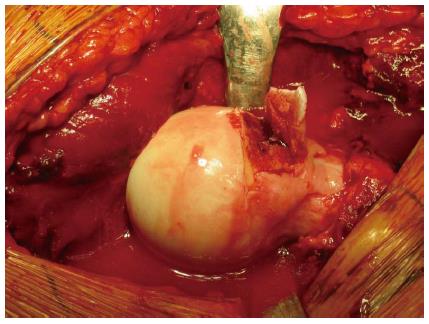
Figure 3 Impingement at the femoral head neck junction is corrected using a curved osteotome to shave slivers of the area of concern.
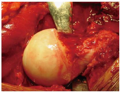
Figure 4 Osteochondroplasty is continued until the femoral head neck junction has been restored to the appropriate anatomical shape to relieve the impingement.
Figure 5 After adequate osteochondroplasty of the femoral head and neck junction and the acetabulum, the labrum is reattached using suture anchors, the femoral head is re-located.
The hip should once again be taken through a full range of motion paying special attention to those positions that were noted to cause impingement prior to dislocation. If adequate resection of the offending lesions has been performed the hip should be able to be taken through a full range of motion with no further impingement.
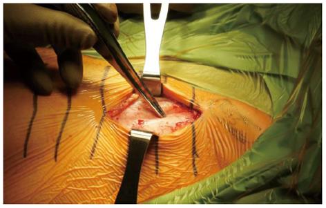
Figure 6 Mini-open technique using “Heuter Approach”, also called the “Short Smith-Pete” because it follows the interval of the Smith-Petersen distal to the anterior superior iliac spine, is used for access to the capsule and femoral neck.
The forceps point to the internervous plane between the femoral nerve (Sartorius) and the superior gluteal nerve (tensor fascia lata).
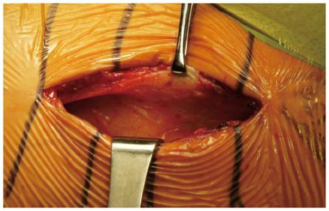
Figure 7 Using “Heuter Approach”, also called the “Short Smith-Pete”, the superficial internervous plane between the femoral nerve (Sartorius) and the superior gluteal nerve (tensor fascia lata) is developed.
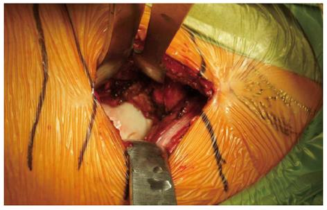
Figure 8 Anterior capsulotomy is performed.
Labrum and femoral head and neck junction is adequately exposed. The distinct inflammatory appearance with red color of the cartilage is visualized.
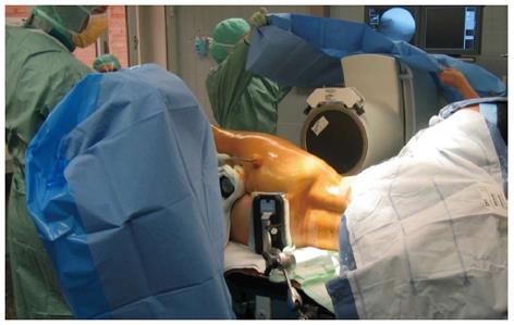
Figure 9 For arthroscopic debridement of femoroacetabular impingement, the patient is placed in a lateral position.
The operative leg is placed in a traction device and fluoroscopy is used to identify the areas of impingement and placement of instruments.
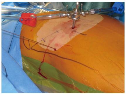
Figure 10 Arthroscopic portal placement: the anterolateral portal is placed about 1 cm proximal and 1 cm anterior to the tip of the greater trochanter.
The anterior portal is placed directly distal to the anterosuperior iliac spine and medial to the anterolateral portal.
- Citation: Wilson AS, Cui Q. Current concepts in management of femoroacetabular impingement. World J Orthop 2012; 3(12): 204-211
- URL: https://www.wjgnet.com/2218-5836/full/v3/i12/204.htm
- DOI: https://dx.doi.org/10.5312/wjo.v3.i12.204


