Copyright
©2011 Baishideng Publishing Group Co.
World J Orthop. Nov 18, 2011; 2(11): 102-106
Published online Nov 18, 2011. doi: 10.5312/wjo.v2.i11.102
Published online Nov 18, 2011. doi: 10.5312/wjo.v2.i11.102
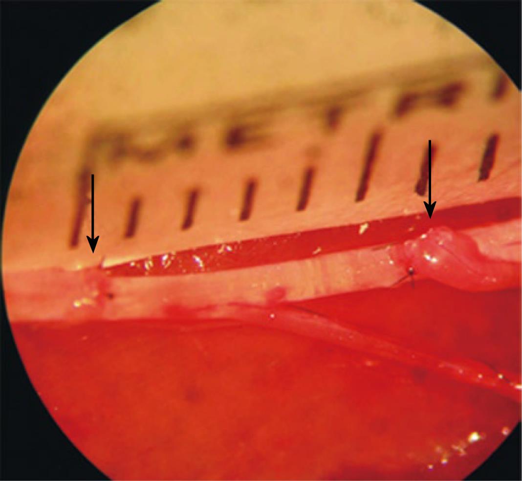
Figure 1 Double end-to-side neurorrhaphy with 0.
6-cm regeneration distance between the proximal and distal stump of the recipient nerve (black arrows). In both neurorrhaphies, coaptation was performed with 3 interrupted 9-0 nylon sutures placed at 120°.
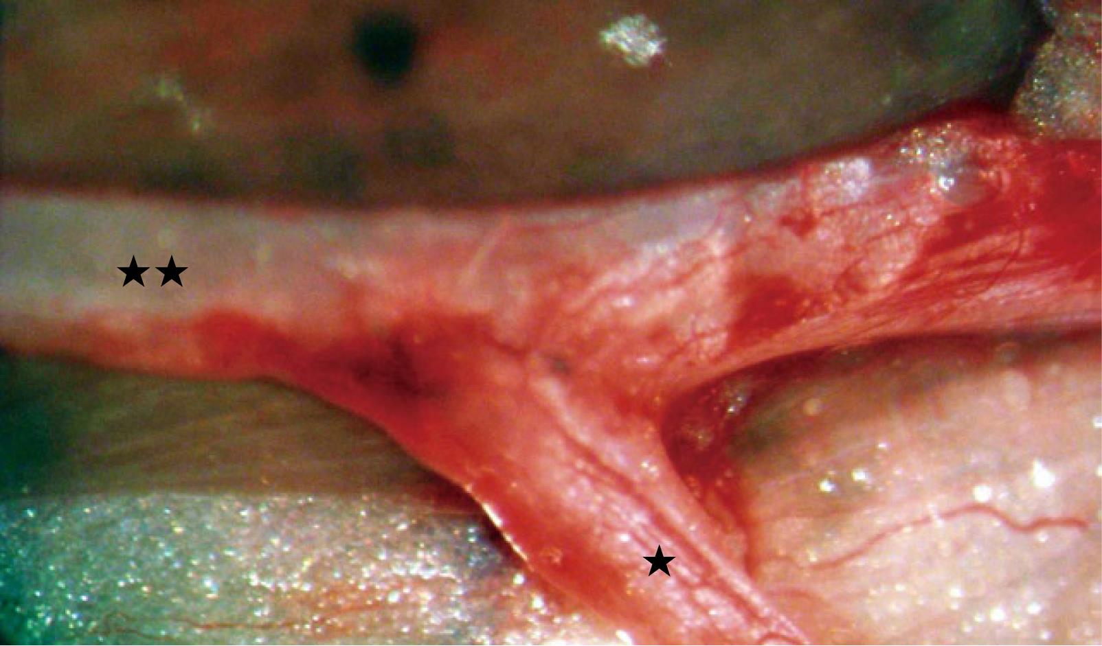
Figure 2 End-to-side neurorrhaphy between the tibial nerve (double asterisk) and the peripheral stump of the peroneal nerve (single asterisk) 90 days after surgery.
Note the smooth transition from one trunk to the other resembling normal bifurcation of the tibial nerve. Also note the newly formed vessels at the outer layer of the nerve trunks traveling from the donor tibial nerve to the recipient peroneal nerve.
- Citation: Lykissas MG. Current concepts in end-to-side neurorrhaphy. World J Orthop 2011; 2(11): 102-106
- URL: https://www.wjgnet.com/2218-5836/full/v2/i11/102.htm
- DOI: https://dx.doi.org/10.5312/wjo.v2.i11.102


