Copyright
©The Author(s) 2025.
World J Orthop. Jul 18, 2025; 16(7): 107045
Published online Jul 18, 2025. doi: 10.5312/wjo.v16.i7.107045
Published online Jul 18, 2025. doi: 10.5312/wjo.v16.i7.107045
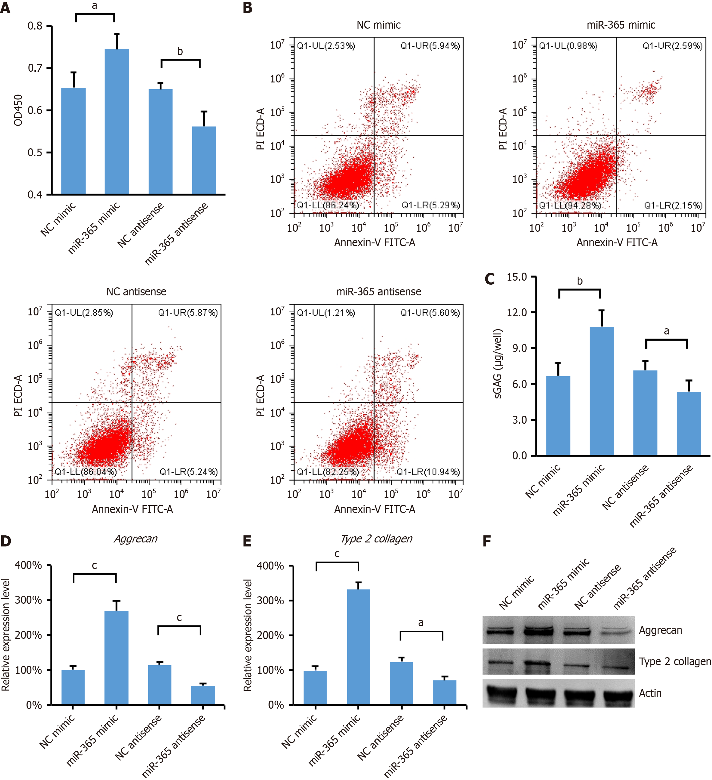
Figure 1 miR-365 promotes differentiation of mesenchymal stem cells into nucleus pulposus cells.
A: CCK-8 assay showed that miR-365 overexpression significantly increased the ability of mesenchymal stem cell (MSC) proliferation, and downregulation of miR-365 showed an inverse effect (n = 4 per group); B: Flow cytometry analysis of annexin-V and propidium iodide staining of apoptotic MSCs with miR-365 overexpression or downregulation; C: Glycosaminoglycans of MSCs were determined in MSC and nucleus pulposus cell (NPC) co-culture system (n = 4 per group); D and E: The relative mRNA level of aggrecan (D) and type 2 collagen (E) from MSCs were determined in MSC and NPC co-culture system (n = 3 per group); F: The protein levels of aggrecan and type 2 collagen from MSCs were determined by Western blot. Data are presented as the mean ± SD. aP < 0.001, bP < 0.01, and cP < 0.05; NC: Negative control.
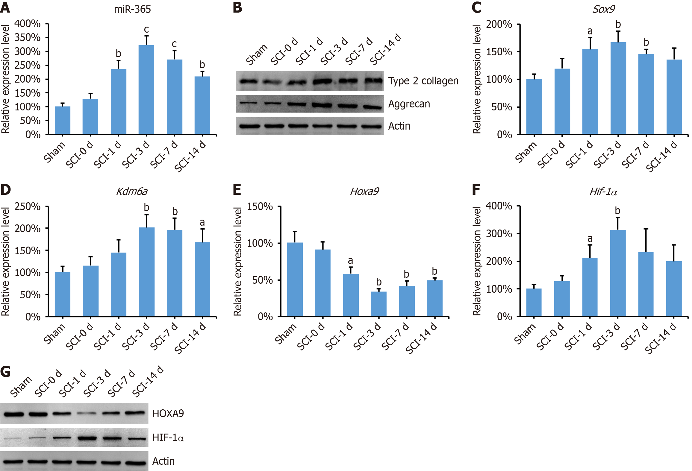
Figure 2 The expression level of miR-365 is increased in the spinal cord of spinal cord injury rats.
A: Relative expression level of miR-365 in the spinal cord of spinal cord injury (SCI) rats (n = 3 per group); B: Relative protein level of aggrecan and type 2 collagen in the spinal cord of SCI rats; C-F: Relative mRNA level of Sox9 (C), Kdm6aA (D), Hoxa9 (E), and Hif-1α (F) in the spinal cord of SCI rats (n = 3 per group); G: Protein level of HOXA9 and HIF-1α in the spinal cord of SCI rats. Data are presented as the mean ± SD. aP < 0.001, bP < 0.01, and cP < 0.05 compared to the sham group.
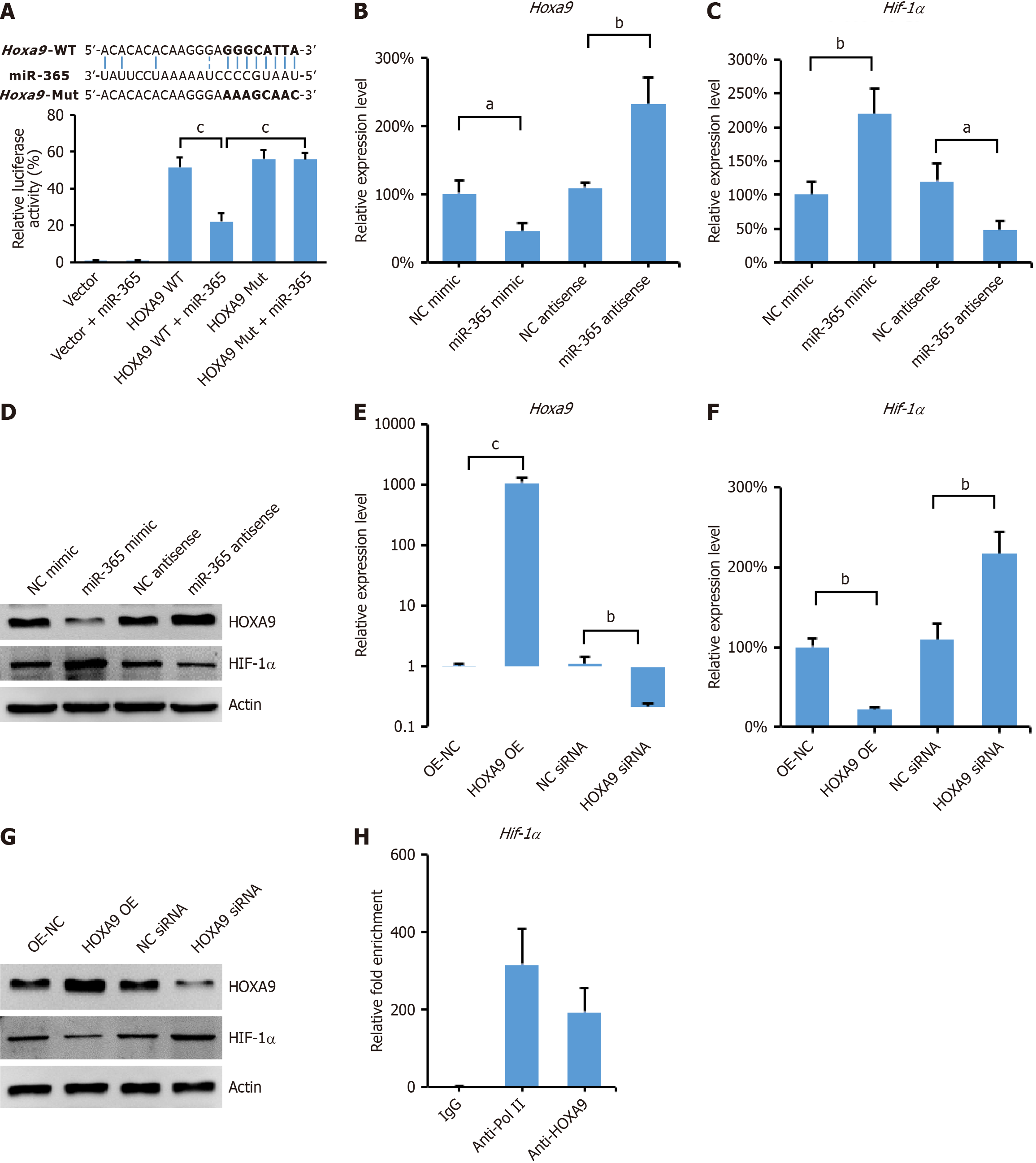
Figure 3 miR-365 binds 3’-UTR of Hoxa9, contributing to increased level of HIF-1α.
A: Luciferase activity assay in mesenchymal stem cells (MSCs) expressing HOXA9-3’UTR-WT with miR-365 mimic or Hoxa9-3’UTR-Mut with miR-365 mimic (n = 4 per group); B and C: Relative mRNA level of Hoxa9 (B) and Hif-1α (C) in MSCs transfected with miR-365 mimic or antisense inhibitor (n = 3 per group); D: Relative protein level of HOXA9 and HIF-1α in MSCs transfected with miR-365 mimic or antisense inhibitor; E and F: Relative mRNA level of Hoxa9 (E) and Hif-1α (F) in MSCs transfected with Hoxa9 plasmid or siRNA (n = 3 per group); G: Protein level of HOXA9 and HIF-1α in MSCs transfected with Hoxa9 plasmid or siRNA; H: Quantitative PCR analyses of chromatin immunoprecipitation assay of HOXA9 binding to the Hif-1α promoter in MSCs (n = 3 per group). Data are presented as the mean ± SD. aP < 0.001, bP < 0.01, and cP < 0.05.
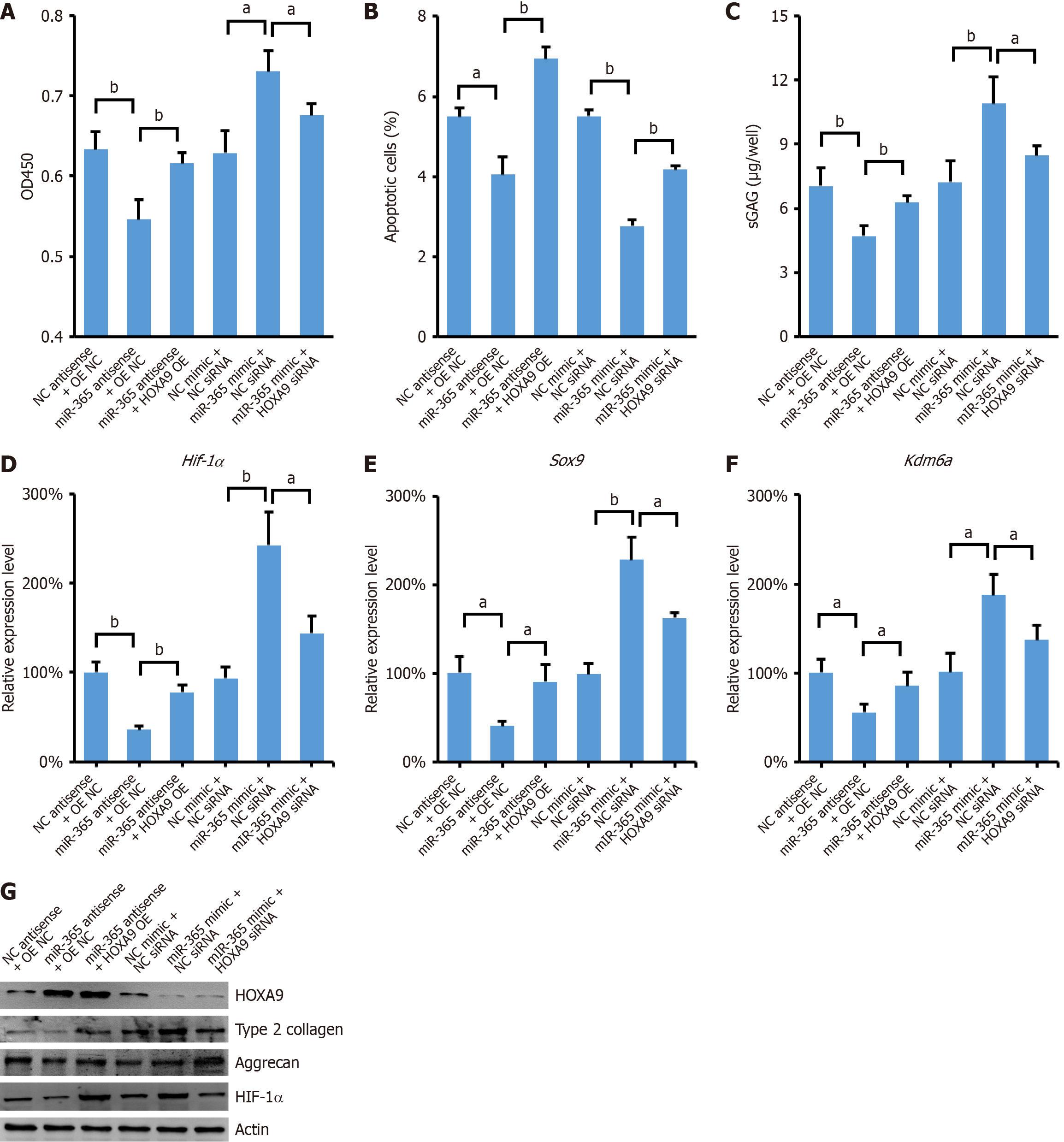
Figure 4 miR-365 regulates differentiation of mesenchymal stem cells into nucleus pulposus cells through HOXA9/HIF-1α axis.
A: Determination of proliferation ability of mesenchymal stem cells (MSCs) with indicated treatment (n = 4 per group); B: Apoptotic MSCs detected by annexin V-FITC/PI staining (n = 3 per group); C: GAGs of MSCs were determined in MSC and nucleus pulposus cell (NPC) co-culture system (n = 4 per group); D-F: The relative mRNA level of Hif-1α (D), Sox9 (E), and Kdm6a (F) in MSCs were determined in MSC and NPC co-culture system (n = 3 per group); G: The indicated protein levels in MSCs were determined by Western blot. Data are presented as the mean ± SD. aP < 0.001, bP < 0.01, and cP < 0.05.
- Citation: Zhi ZZ, Liu T, Kang J, Zhou FC, Liu XD, He ZM. miR-365 promotes HOXA9-mediated differentiation of mesenchymal stem cells to nucleus pulposus cells by interacting with HIF-1α. World J Orthop 2025; 16(7): 107045
- URL: https://www.wjgnet.com/2218-5836/full/v16/i7/107045.htm
- DOI: https://dx.doi.org/10.5312/wjo.v16.i7.107045


