Copyright
©The Author(s) 2025.
World J Orthop. Jul 18, 2025; 16(7): 105111
Published online Jul 18, 2025. doi: 10.5312/wjo.v16.i7.105111
Published online Jul 18, 2025. doi: 10.5312/wjo.v16.i7.105111
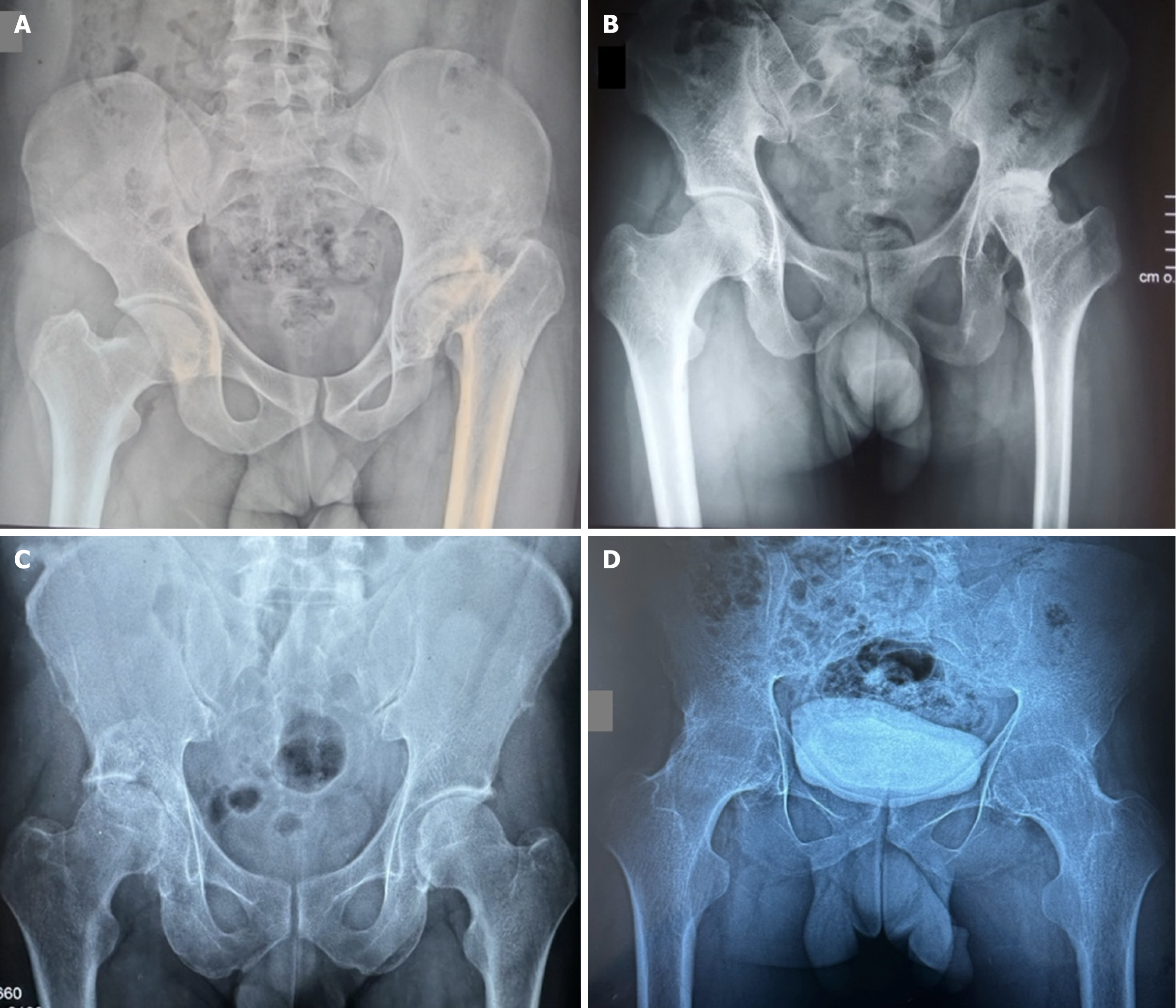
Figure 1 Radiographic images illustrating the various pathologies.
A: Sequela of epiphysiolysis of the right hip; B: Sequela of right Legg-Calvé-Perthes disease; C: Post-traumatic left coxarthrosis following a fracture of the acetabular roof; D: Bilateral ankylosis of the hip.
Figure 2 Disarthrodesis-prosthesis.
A: Evidence of fusion of the femoral head in the acetabulum, after osteotomy of the neck; B: Milling of the head; C: Placement of the prosthetic acetabulum; D: Posterior.
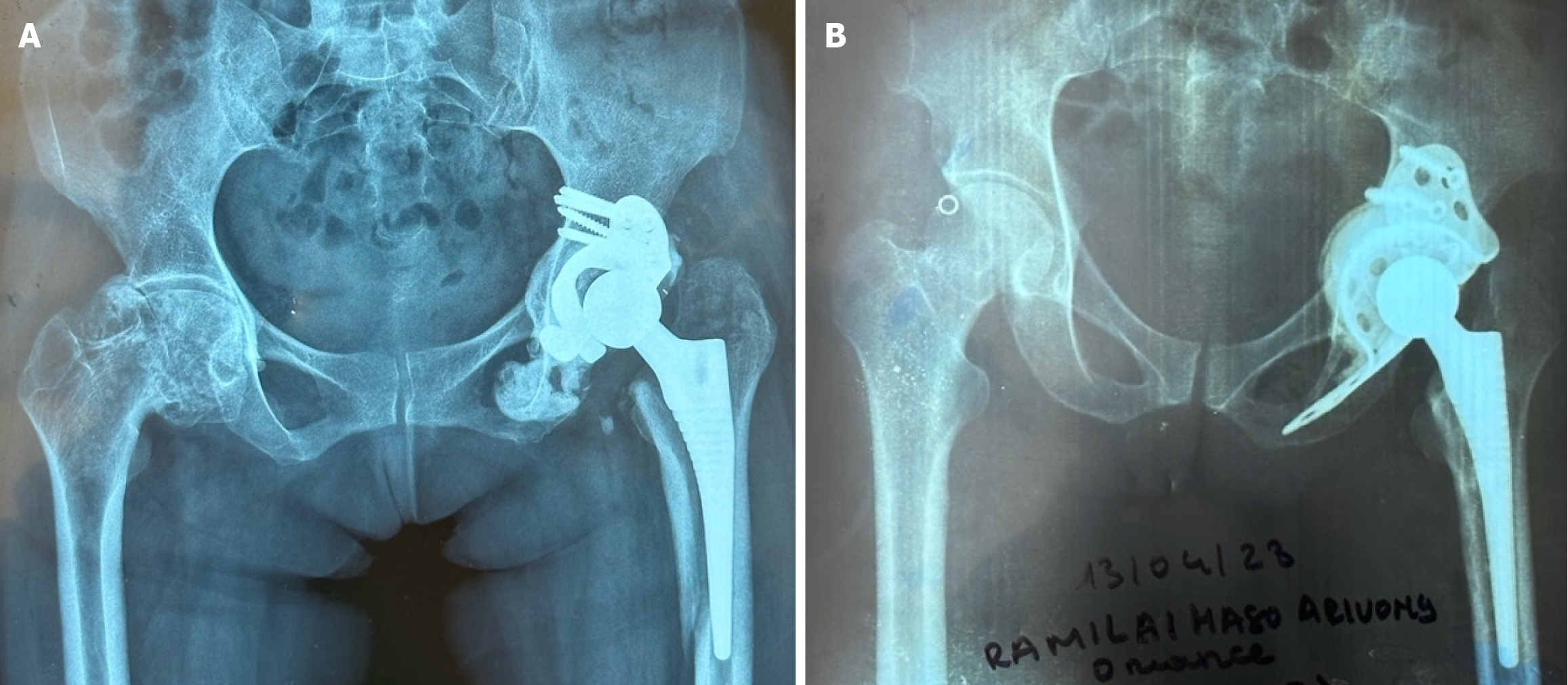
Figure 3 Acetabular reconstruction.
A: Kerboull support ring; B: Bürch-Schneider anti-protrusion cage.
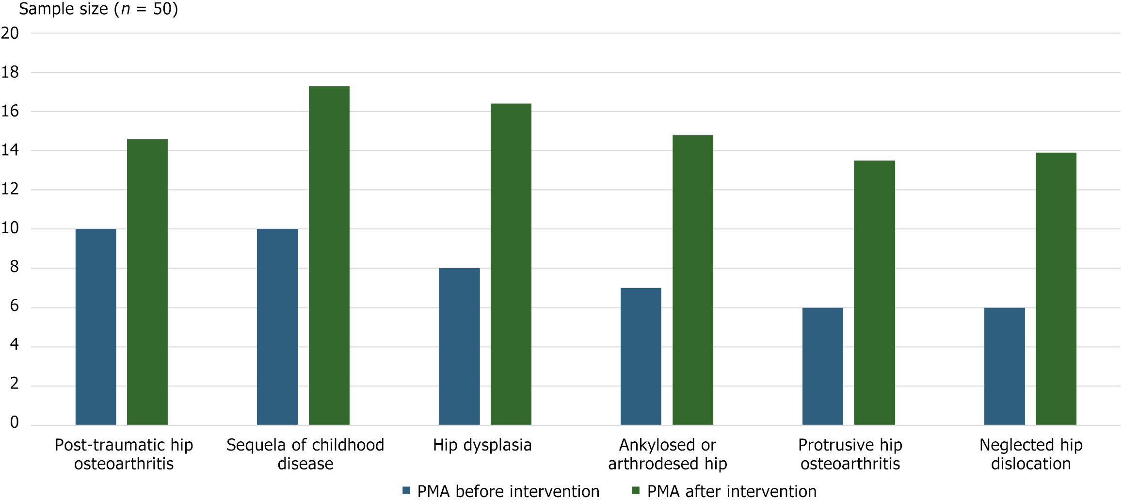
Figure 4 Postel Merle d'Aubigné score distribution before and after surgery.
PMA: Postel Merle d'Aubigné.
- Citation: Manasse H, Daoulas T, Rohimpitiavana AS, Solofomalala GD, Dubrana F, Razafimahandry HJC. Surgical techniques and outcomes of difficult total hip replacements: A challenge in a low-income country. World J Orthop 2025; 16(7): 105111
- URL: https://www.wjgnet.com/2218-5836/full/v16/i7/105111.htm
- DOI: https://dx.doi.org/10.5312/wjo.v16.i7.105111


