Copyright
©The Author(s) 2025.
World J Orthop. Feb 18, 2025; 16(2): 98674
Published online Feb 18, 2025. doi: 10.5312/wjo.v16.i2.98674
Published online Feb 18, 2025. doi: 10.5312/wjo.v16.i2.98674
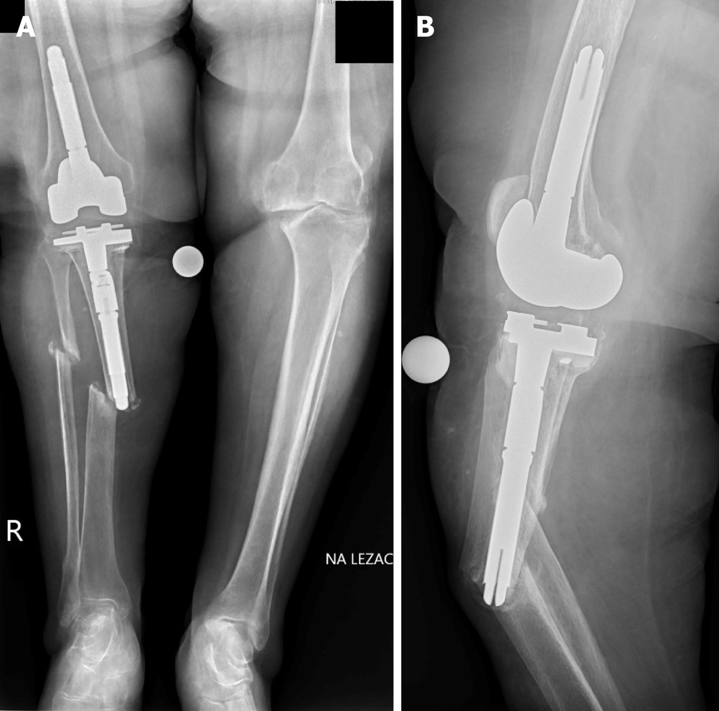
Figure 1 X-ray images of a 65-year-old woman with periprosthetic fracture of the right tibia (Felix type 2A) and pseudarthrosis occurrence.
A: Anteroposterior view; B: Lateral view. There were no signs of prosthesis component loosening.
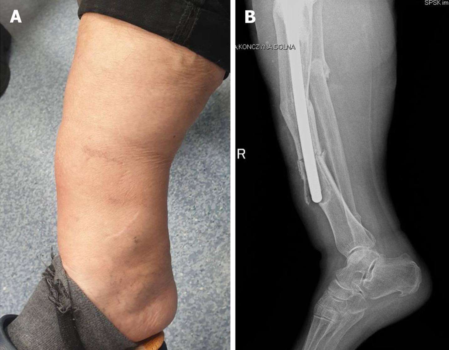
Figure 2 Periprosthetic fracture of the anterior cortex of the tibia at the top of the endoprosthesis stem and another fracture gap at the level of the stem.
A: Deformation and swelling of the distal shin; B: Lateral view radiogram. Endoprosthesis and proximal part of the stem without signs of loosening.
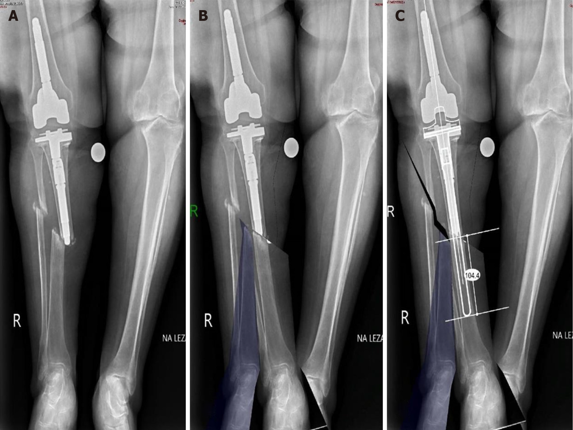
Figure 3 Sequence of preoperative planning and prosthesis digital templating (OrthoViewTM).
A: Before planning; B: Reduction; C: Prosthesis with long stem templating. A radiograph with matching of the long-stemmed tibial component, fracture reduction, and correction of limb axis. The tip of the stem exceeds 104 mm over the fracture level.
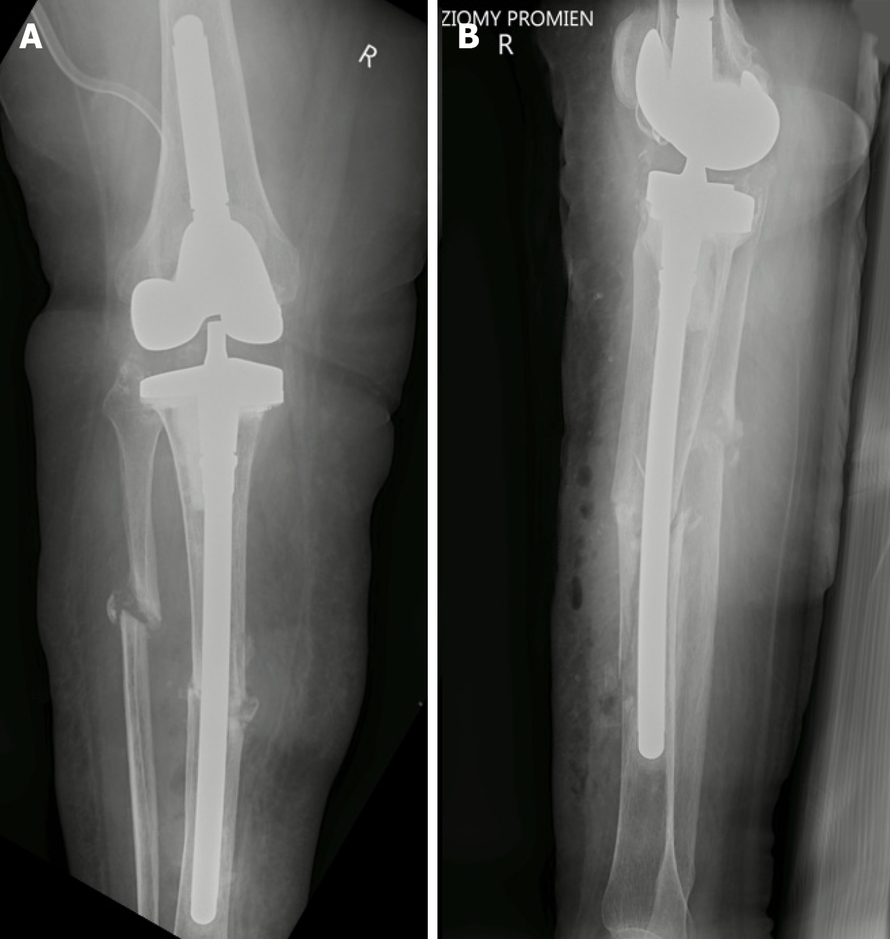
Figure 4 Post-operative radiographs confirmed the correct position of the tibial implant with fracture reduction and stabilization on a long, uncemented prosthesis stem.
A: Anteroposterior view; B; Lateral view.
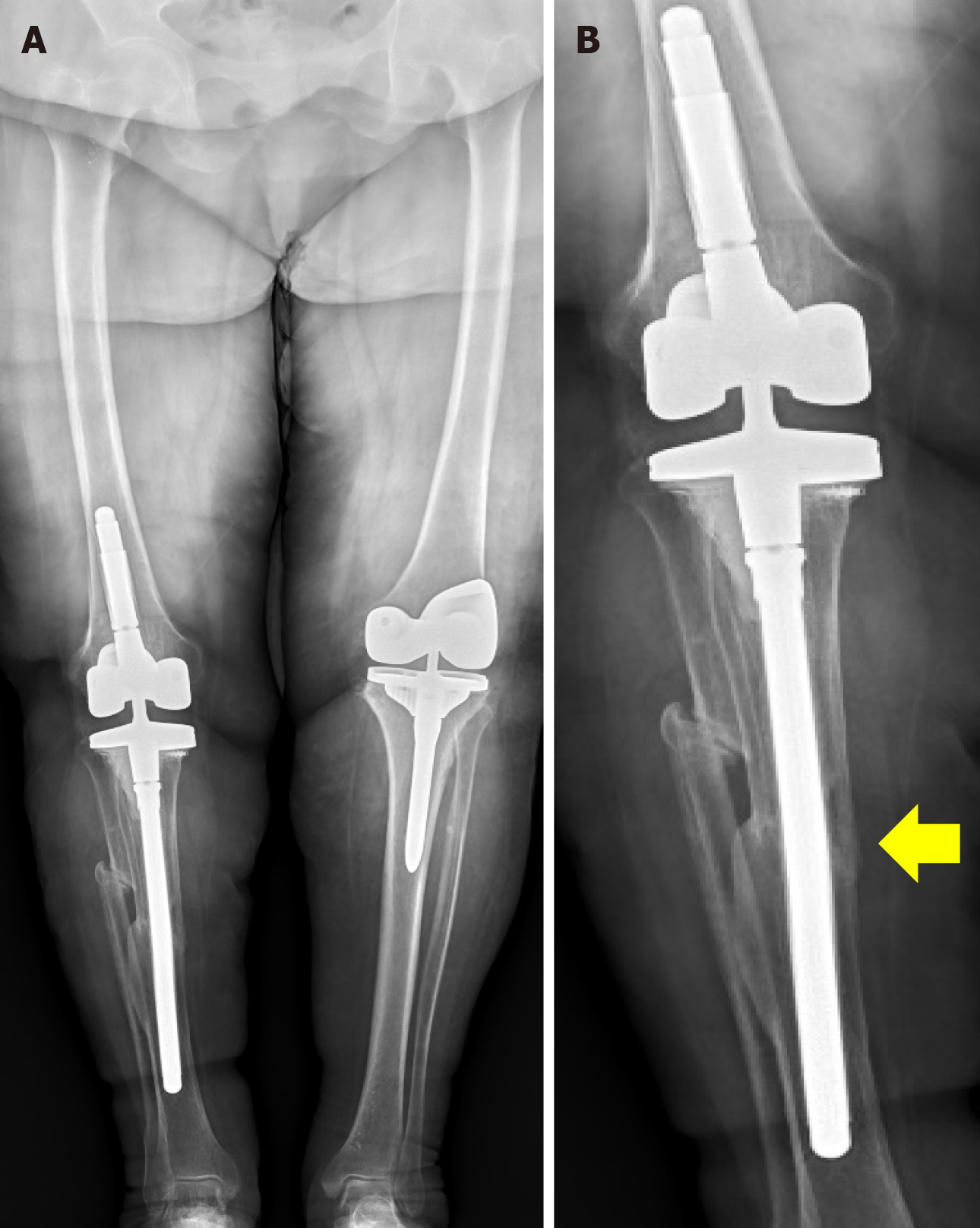
Figure 5 Radiographs 2 years after revision total knee arthroplasty of the right knee, and 6 months after left total knee arthroplasty.
A: Standing long leg; B: Plain anteroposterior. Full bone union of the right tibia was confirmed (yellow arrow). Distally, progressive loss of the anterior tibial cortex was revealed as a result of the load exerted by the distal part of stem.
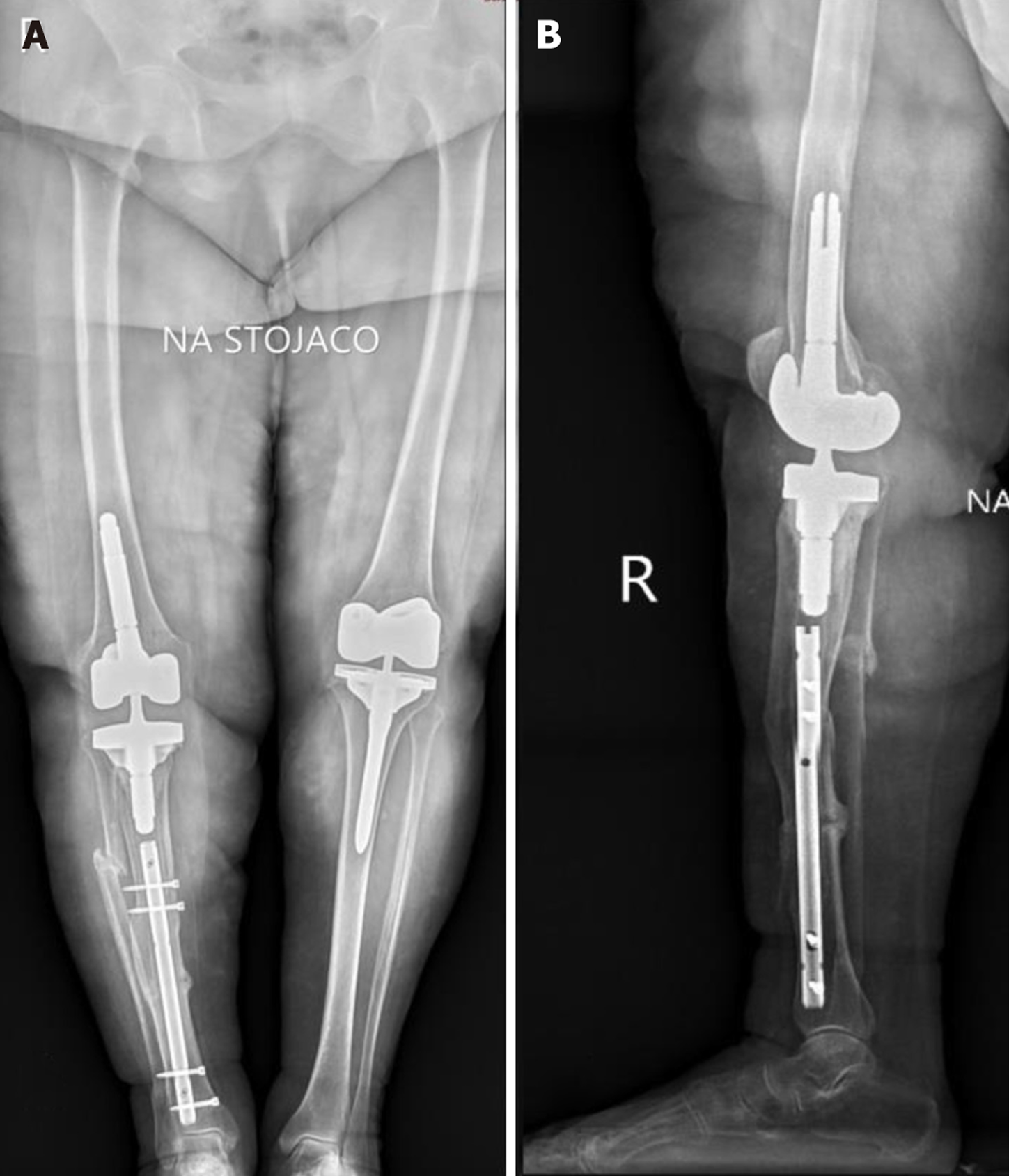
Figure 6 Post-operative radiographs confirmed the correct position of the tibial implant and intramedullary nails with proper fracture reduction and stabilization.
A: Anteroposterior view; B: Lateral view.
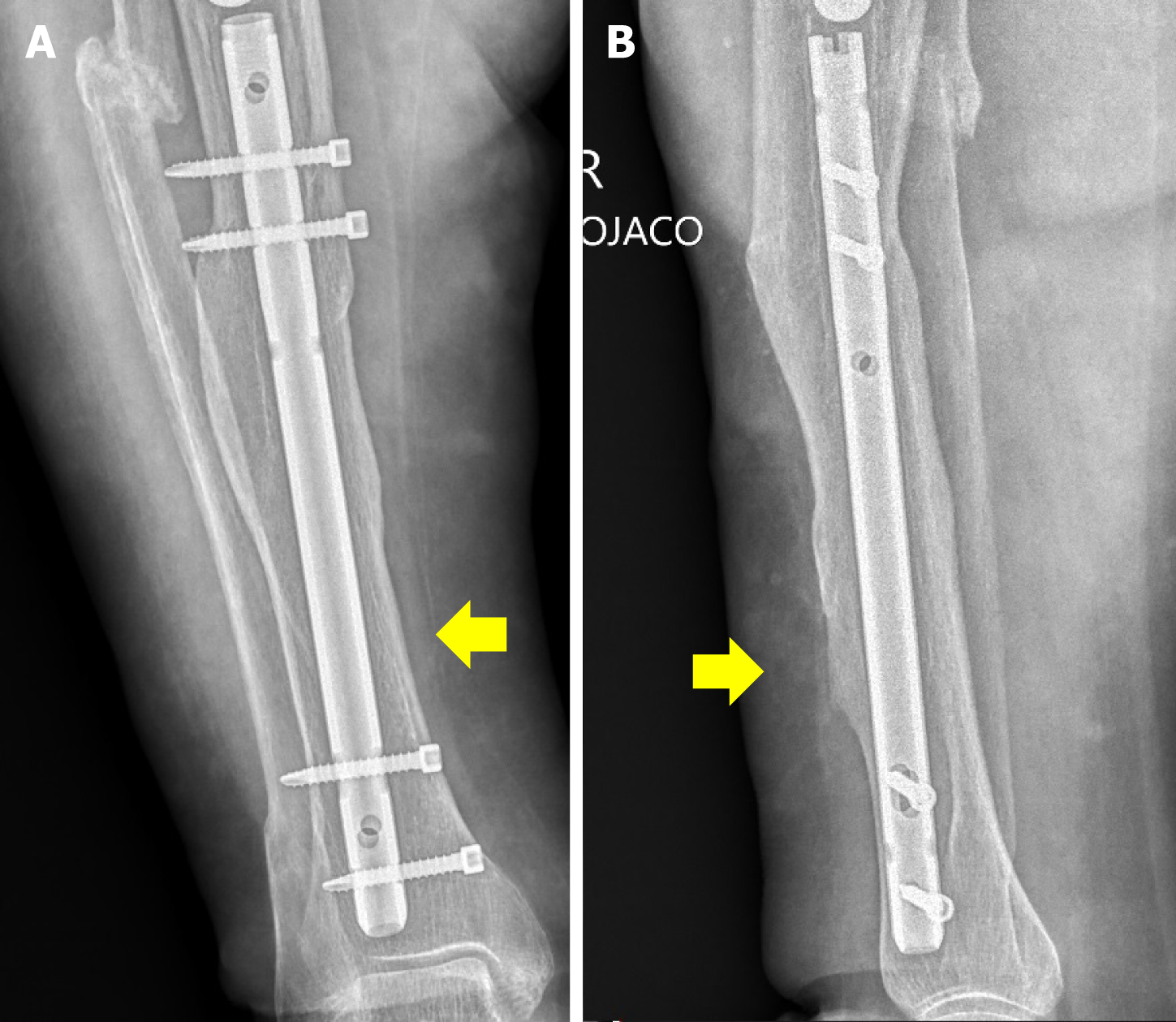
Figure 7 At the last follow-up visit 26 months after surgery, full bone union was confirmed.
A: Anteroposterior view; B: Lateral view.
- Citation: Kocon M, Grzelecki D. Periprosthetic fractures of the tibial shaft following long-stemmed total knee arthroplasty: A case report. World J Orthop 2025; 16(2): 98674
- URL: https://www.wjgnet.com/2218-5836/full/v16/i2/98674.htm
- DOI: https://dx.doi.org/10.5312/wjo.v16.i2.98674


