Copyright
©The Author(s) 2025.
World J Orthop. Feb 18, 2025; 16(2): 104095
Published online Feb 18, 2025. doi: 10.5312/wjo.v16.i2.104095
Published online Feb 18, 2025. doi: 10.5312/wjo.v16.i2.104095
Figure 1 The computed tomography and magnetic resonance imaging images of the patient.
A: There are degenerative changes in the cervical spine, with a herniated disc at C3/4; B and C: The cervical spine computed tomography scans show that the odontoid is located 7.32 mm above the chamberlain line, suggesting basilar invagination and compression of the cervical spinal cord.
Figure 2 Cervical dynamic X-ray examination suggests instability at C1/2.
A: The active dose indicator (ADI) measures 9.49 mm in extension; B: The ADI measures 5.36 mm in the neutral position, 9.49 mm in extension; C: The ADI measures 10.75 mm in flexion, diagnosing atlantoaxial dislocation.
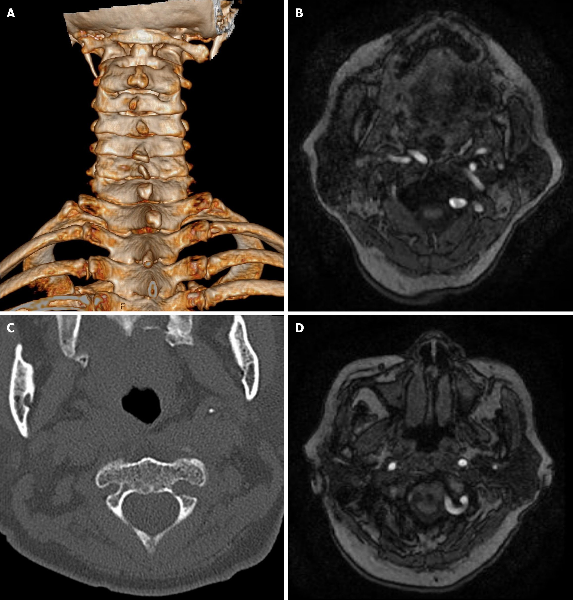
Figure 3 Preoperative computed tomography reconstruction and magnetic resonance tomography images of the patient.
A: Computed tomography (CT) reconstruction scans demonstrated spontaneous atlanto-occipital fusion; B-D: CT cross-sectional images indicate a high-riding vertebral artery anomaly and restricted placement of screws on the left side of the C2 vertebra. Vascular MRI imaging indicates invasion of the left vertebral artery into the lateral mass of C2 and the left posterior arch of C1.
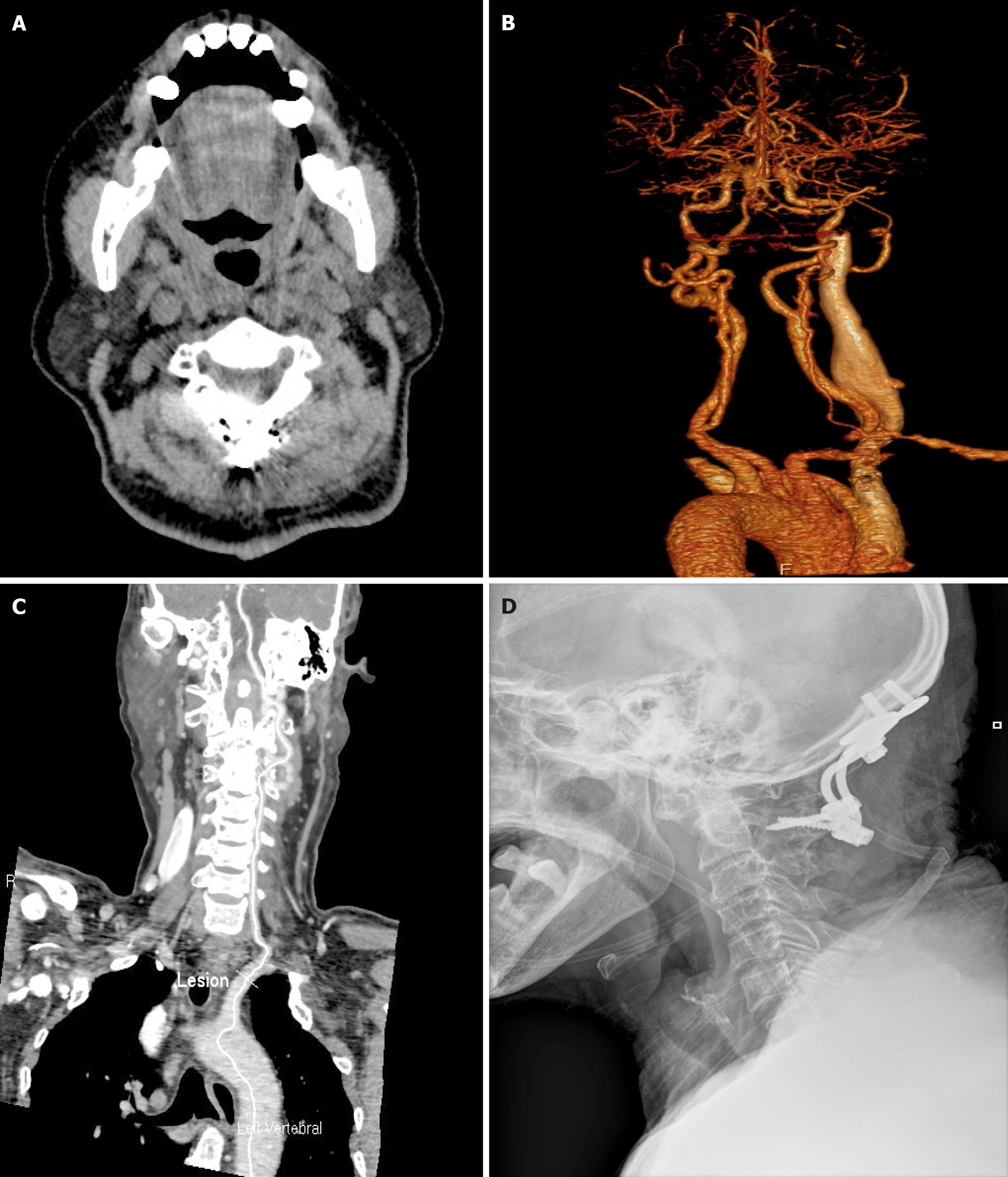
Figure 4 Images of the patient after surgery and during follow-up.
A: Postoperative cervical computed tomography (CT) scans demonstrated screws in situ within the C2 Lamina; B and C: Postoperative CT angiography showed a patent left vertebral artery without post-traumatic stenosis; D: Postoperative radiographs revealed the internal fixation to be well-positioned.
- Citation: Tang RH, Yin J, Zhou ZW. Atlantoaxial dislocation with vertebral artery anomaly: A case report. World J Orthop 2025; 16(2): 104095
- URL: https://www.wjgnet.com/2218-5836/full/v16/i2/104095.htm
- DOI: https://dx.doi.org/10.5312/wjo.v16.i2.104095


