Copyright
©The Author(s) 2024.
World J Orthop. Aug 18, 2024; 15(8): 820-827
Published online Aug 18, 2024. doi: 10.5312/wjo.v15.i8.820
Published online Aug 18, 2024. doi: 10.5312/wjo.v15.i8.820
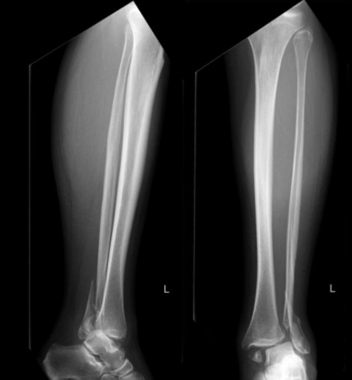
Figure 1 Trimalleolar Weber B fracture with subluxation.
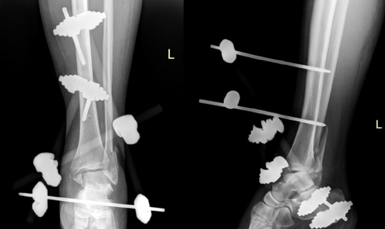
Figure 2 X-ray after closed reduction and external fixation using ex-fix (DePuy Synthes, West Chester, PA, United States).
Two Pins in tibia, one pin in calcaneus.
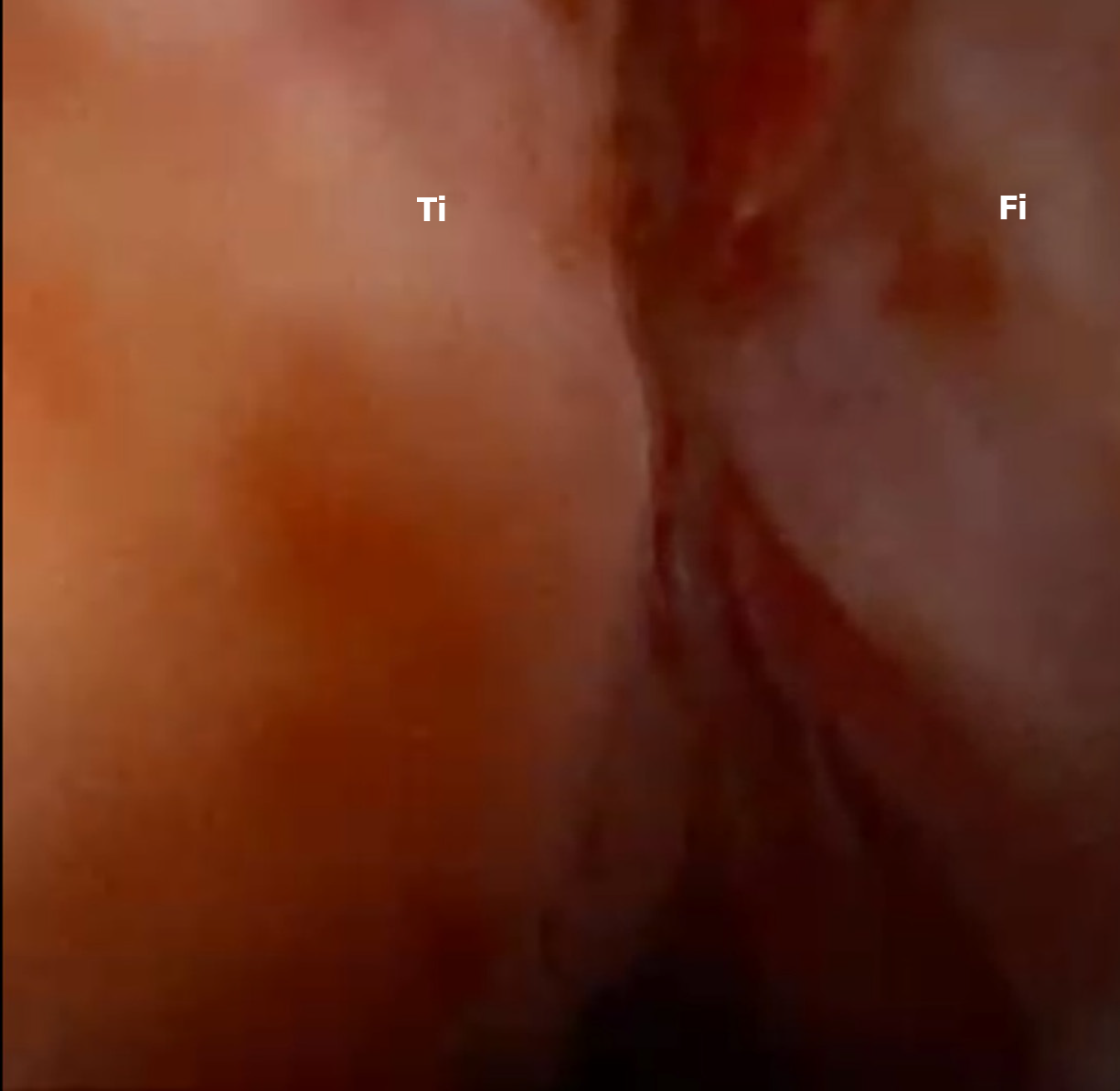
Figure 3 Tibiofibular syndesmosis injury in nanoscopy camera.
Ti: Tibia; Fi: Fibula.
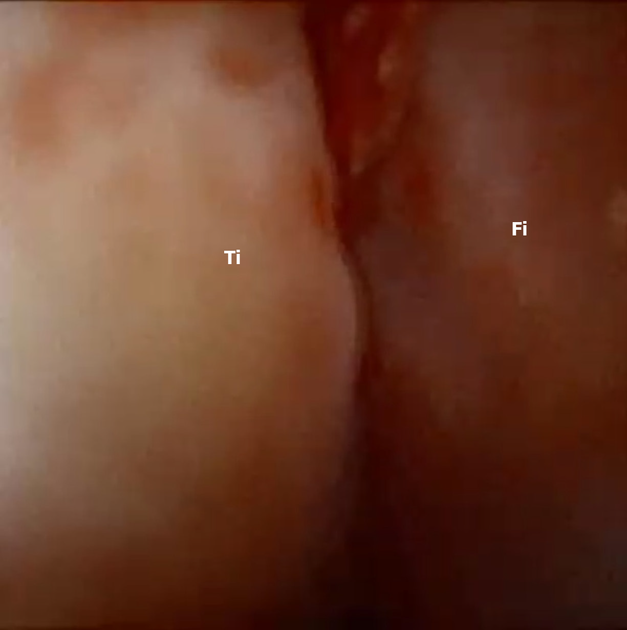
Figure 4 Tibiofibular syndesmosis after repair in nanoscopy camera.
Ti: Tibia; Fi: Fibula.
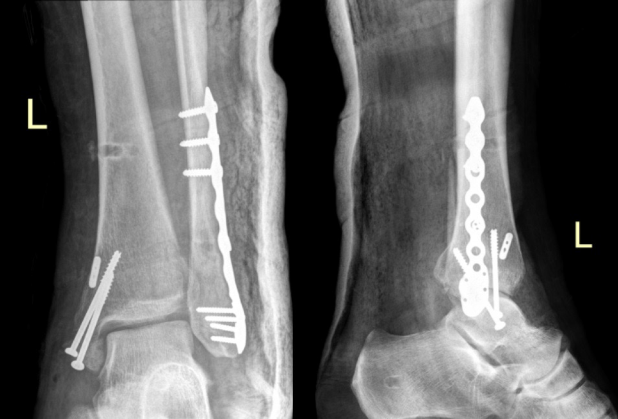
Figure 5 X-ray after open reduction and internal fixation with plate, screws and suture button (Arthrex, Naples, FL, United States).
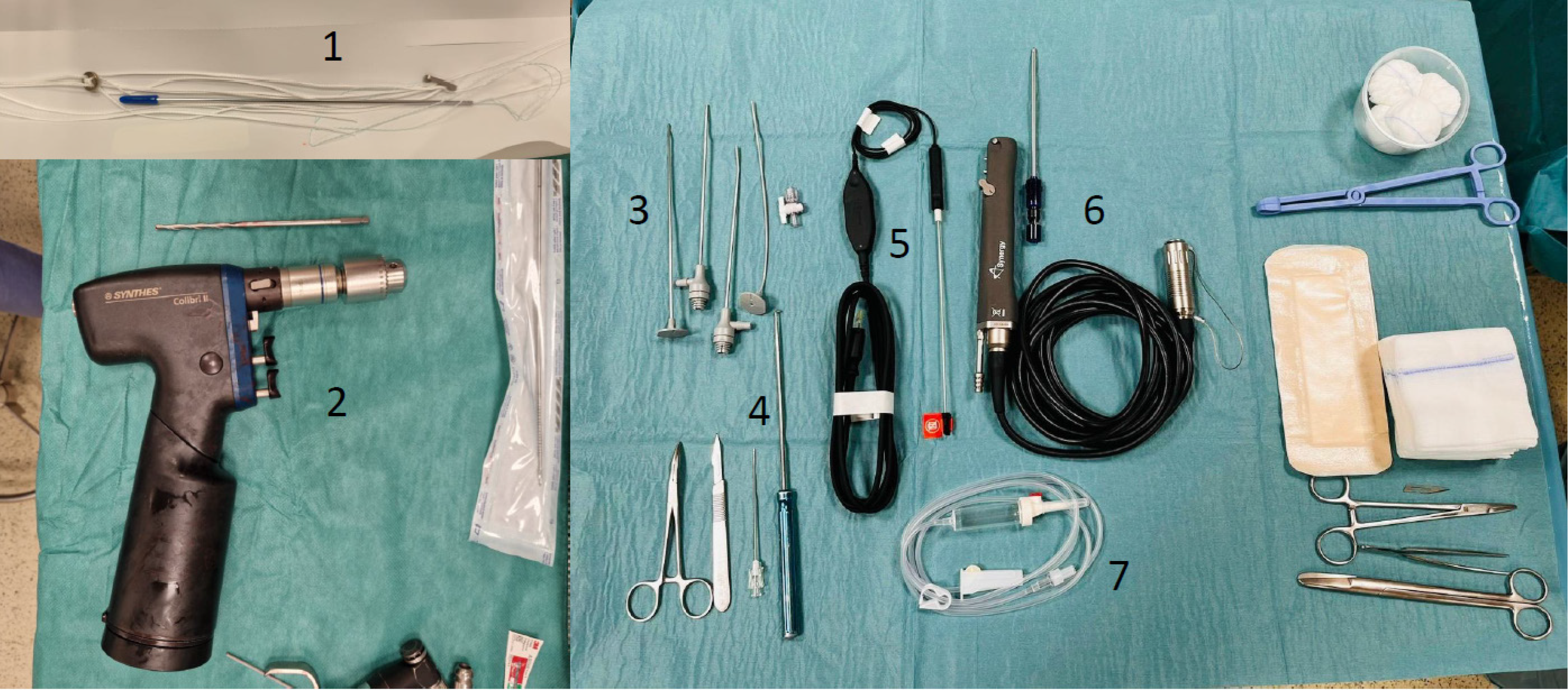
Figure 6 Instruments.
1: Suturebutton; 2: Drill; 3: Trocars; 4: Hook; 5: Nanoscope; 6: Shaver; 7: Water supply.
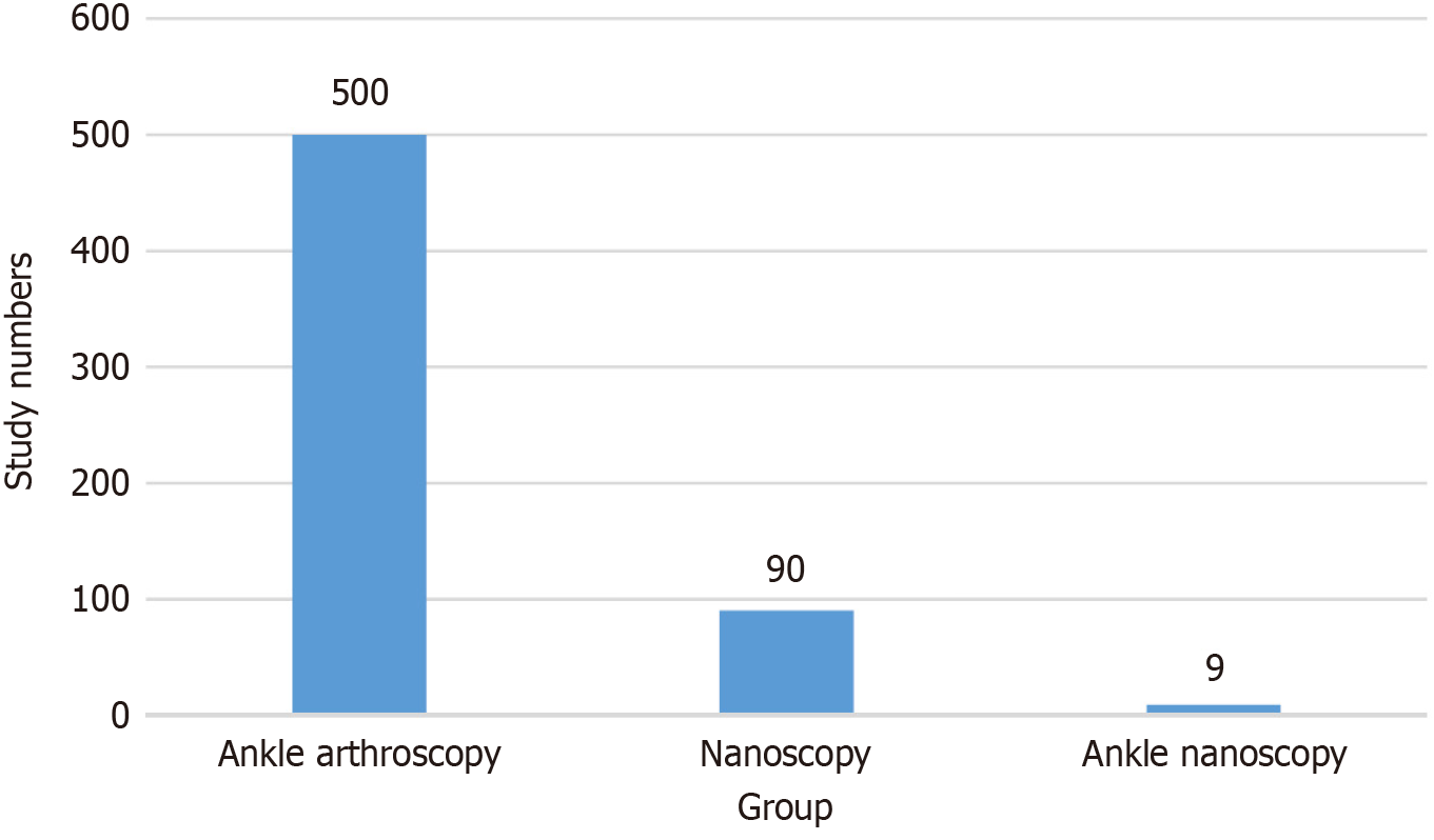
Figure 7 Number of studies about ankle nanoscopy.
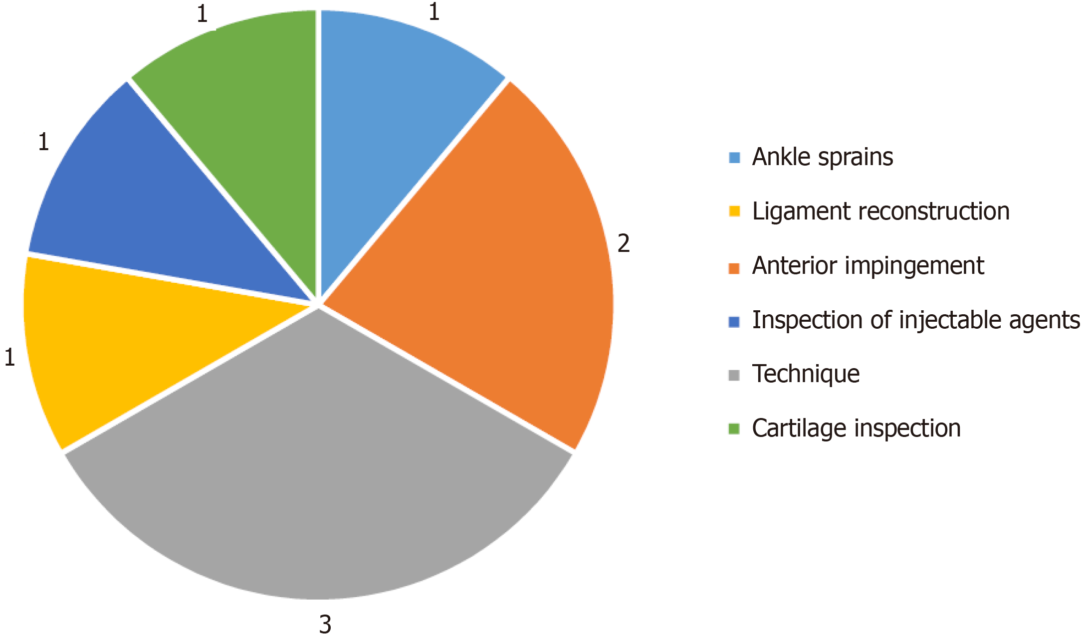
Figure 8 Subjects of studies about ankle nanoscopy.
- Citation: Wojtowicz BG, Domzalski M, Lesman J. Needle arthroscopic-assisted repair of tibio-fibular syndesmosis acute injury: A case report. World J Orthop 2024; 15(8): 820-827
- URL: https://www.wjgnet.com/2218-5836/full/v15/i8/820.htm
- DOI: https://dx.doi.org/10.5312/wjo.v15.i8.820


