Copyright
©The Author(s) 2024.
World J Orthop. Aug 18, 2024; 15(8): 807-812
Published online Aug 18, 2024. doi: 10.5312/wjo.v15.i8.807
Published online Aug 18, 2024. doi: 10.5312/wjo.v15.i8.807
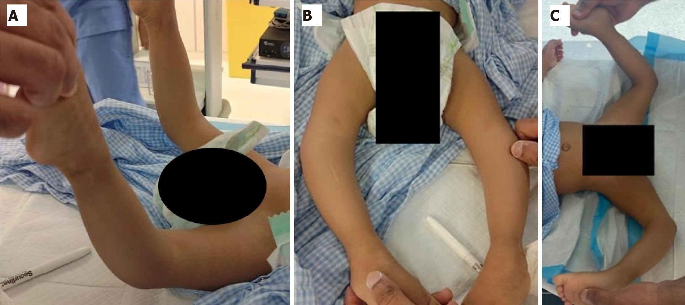
Figure 1 Preoperative examination.
A: Degree of dislocation before the reduction; B: Absence of skin grooves; C: Anterior view before the reduction.
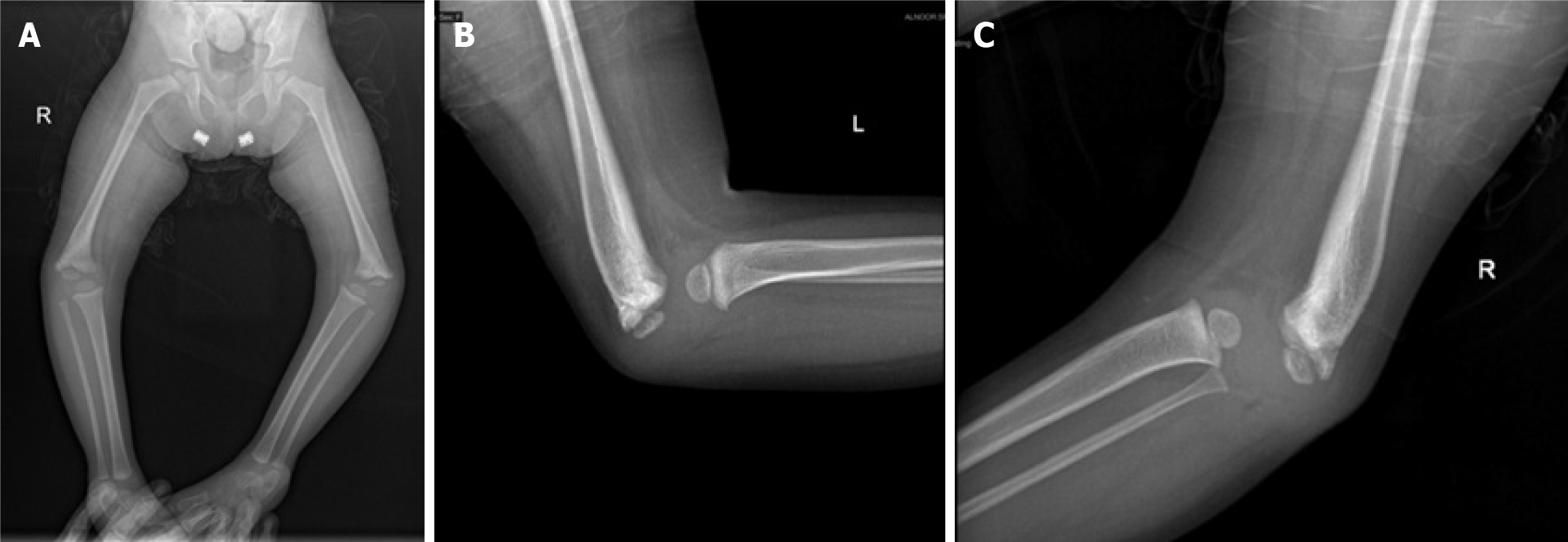
Figure 2 Preoperative radiographic imaging.
A: Anterior-posterior X-ray image of the lower limbs; B: Lateral X-ray view of the left knee; C: Lateral X-ray view of the right knee.
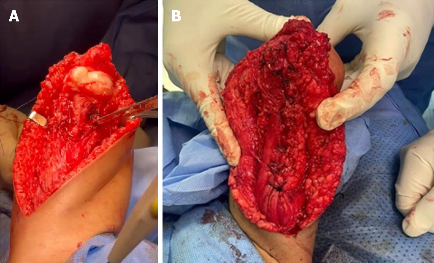
Figure 3 Intraoperative picture.
A: Knee capsule; B: After muscle transfer and before closure of the skin.
Figure 4 Postoperative follow-up.
A: The angle of the knee two months after surgery and after cast removal; B: Four months of surgery, she could walk with assistance and a knee cage to support her knee; C and D: Six months after surgery.
- Citation: Qasim OM, Abdulaziz AA, Aljabri NK, Albaqami KS, Suqaty RM. Neglected congenital bilateral knee dislocation treated by quadricepsplasty with semitendinosus and sartorius transfer: A case report. World J Orthop 2024; 15(8): 807-812
- URL: https://www.wjgnet.com/2218-5836/full/v15/i8/807.htm
- DOI: https://dx.doi.org/10.5312/wjo.v15.i8.807


