Copyright
©The Author(s) 2024.
World J Orthop. Apr 18, 2024; 15(4): 379-385
Published online Apr 18, 2024. doi: 10.5312/wjo.v15.i4.379
Published online Apr 18, 2024. doi: 10.5312/wjo.v15.i4.379
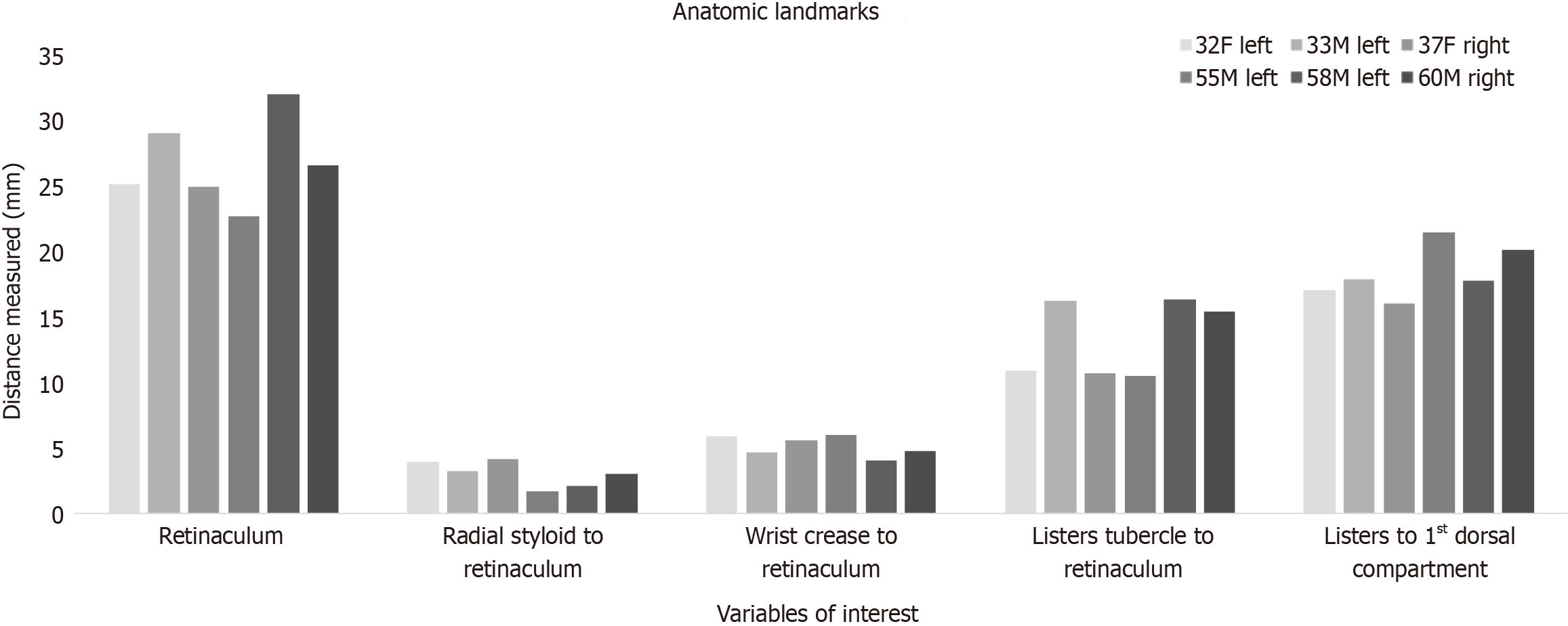
Figure 1 Histogram demonstrating superficial landmark data points for six cadaveric specimens.
Each data bar demonstrates the value obtained for the cadaveric forearm specified in the legend provided. Notably, all variables do not demonstrate a significant outlier and maintain a close distribution of variables independent of laterality, age, and sex of the specimen. This demonstrates that the radial styloid, wrist crease, and Lister’s tubercle are all reliable markers to use to determine the location of the first dorsal compartment.
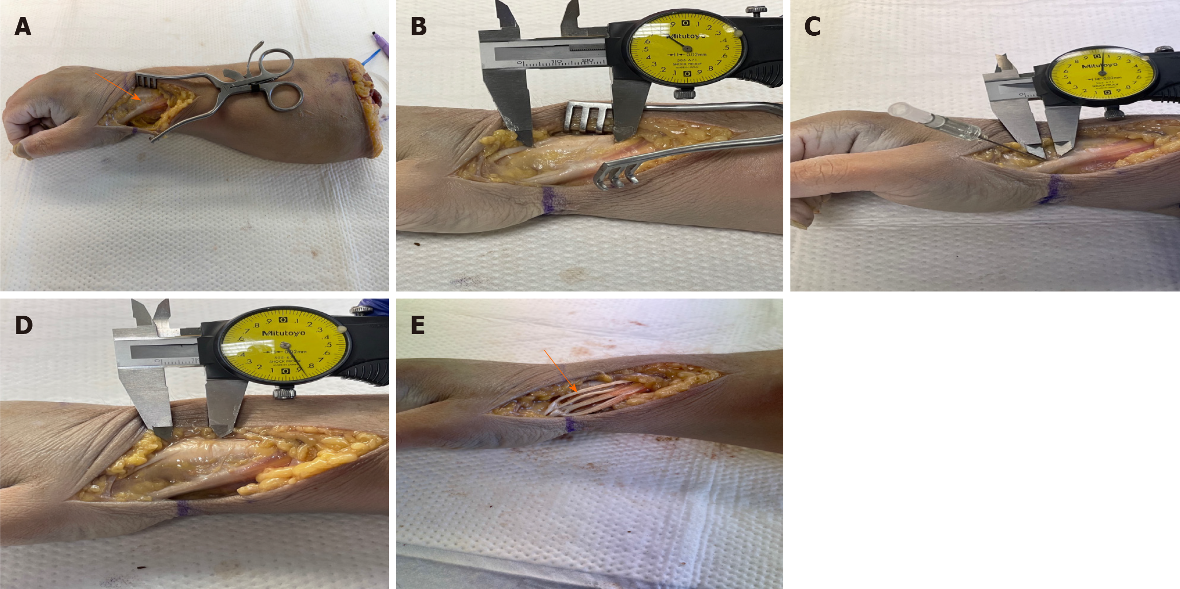
Figure 2 Cadaveric dissection.
A: Initial cadaveric dissection of the first dorsal compartment. Right forearm cadaver model with a longitudinal incision centered over the first dorsal extensor compartment. Superficial dissection through the subcutaneous fat was performed with a scalpel blade and Metzenbaum scissors. Deep dissection and retraction of the soft tissues demonstrates the underlying musculature and extensor retinaculum as shown by the black arrow; B: Full length view of the extensor retinaculum. Cadaveric dissection demonstrating the full extent of the extensor retinaculum. Caliper measurements were performed as demonstrated in this graphic. The average length of the extensor retinaculum from its proximal to its distal length was 26.82 mm ± 3.34 mm; C: Deep dissection of the radial styloid and extensor retinaculum. Dissection demonstrating the most distal aspect of the radial styloid to the most distal aspect of the extensor retinaculum. An 18-gauge needle is used to mark the radial styloid process. The length from the radial styloid to the initial aspect of the extensor retinaculum measured 2.98 mm ± 0.99 mm; D: Anatomic relationship shown between Lister’s tubercle to the first dorsal extensor compartment. Lister’s tubercle is seen marked by the most distal aspect of the caliper, while the first extensor compartment shown by the proximal caliper marker. The distance from Lister’s tubercle to the proximal aspect of the retinaculum measured 13.37 mm ± 2.94 mm while distance from Lister’s tubercle to the start of the first dorsal compartment was 18.43 mm ± 2.01 mm; E: Multiple abductor pollicis longus (APL) tendon slips and sub-sheaths. Deep dissection into the first extensor compartment demonstrates multiple tendon slips of the APL tendon as shown by the black arrow. Four separate tendon slips are shown by the arrow, ultimately resulting in incomplete compartment release if not thoroughly dissected. The average number of APL tendon slips was three. A pseudo-retinaculum was also present in four out of six cadavers.
- Citation: Thandoni A, Yetter WN, Regal SM. Anatomic location of the first dorsal extensor compartment for surgical De-Quervain’s tenosynovitis release: A cadaveric study. World J Orthop 2024; 15(4): 379-385
- URL: https://www.wjgnet.com/2218-5836/full/v15/i4/379.htm
- DOI: https://dx.doi.org/10.5312/wjo.v15.i4.379


