Copyright
©The Author(s) 2024.
World J Orthop. Mar 18, 2024; 15(3): 215-229
Published online Mar 18, 2024. doi: 10.5312/wjo.v15.i3.215
Published online Mar 18, 2024. doi: 10.5312/wjo.v15.i3.215
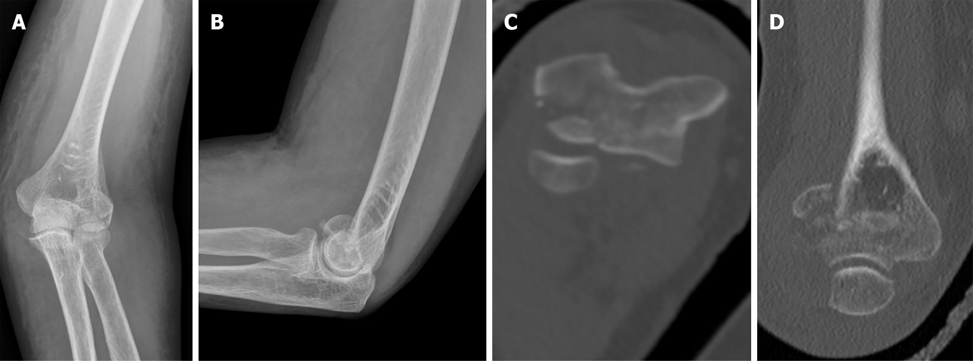
Figure 1 Injury films on presentation depicting a left Bryan and Morrey type 3 capitellar fracture.
A-D: Anterior posterior (A) and lateral (B) radiographs, axial (C) and coronal (D) computer tomography cuts depicting the injury.
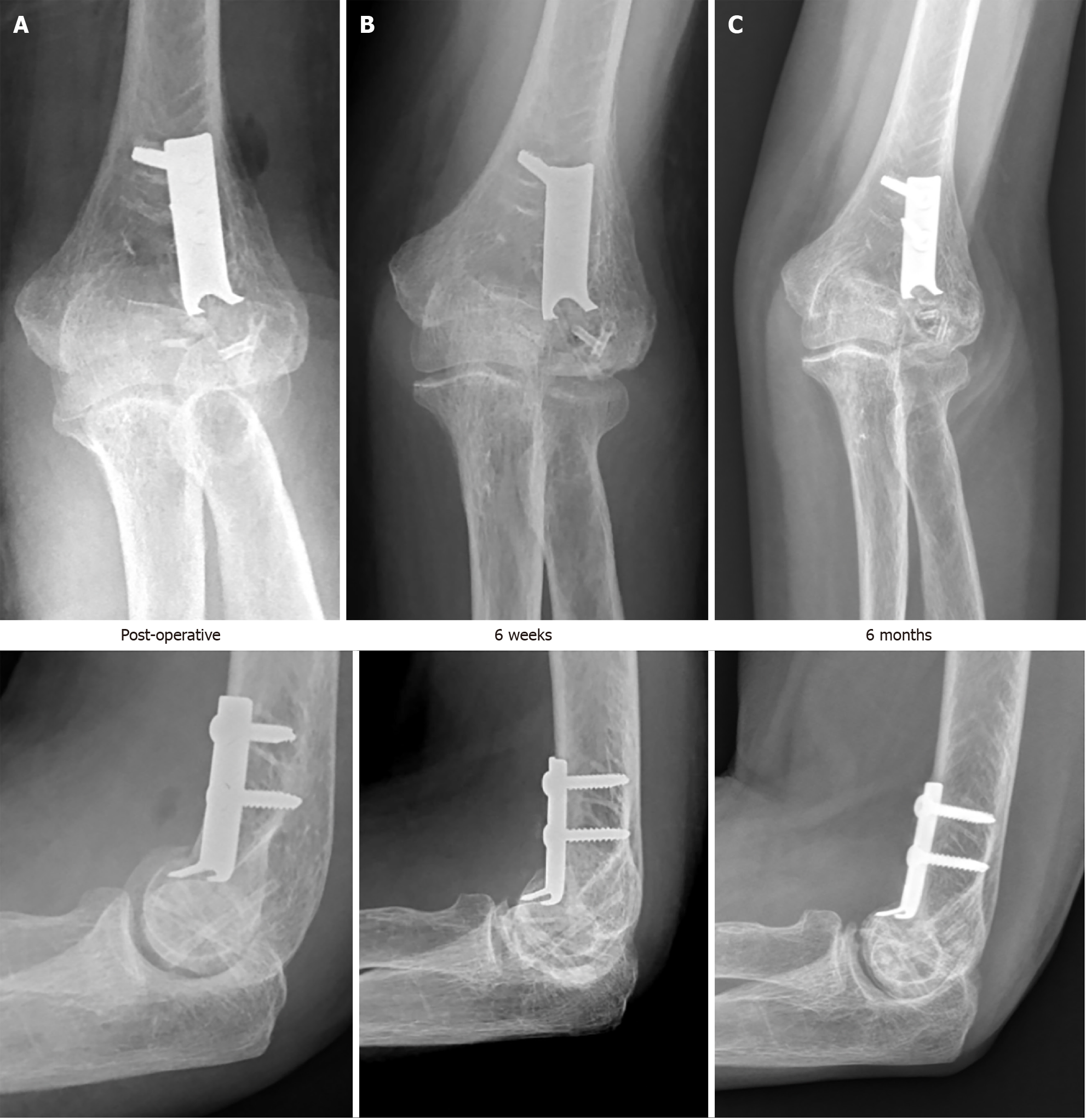
Figure 2 Post operative radiographs depicting progress.
A-C: Radiographs immediately post op (A), at 6 wk (B) and at 6 months (C).
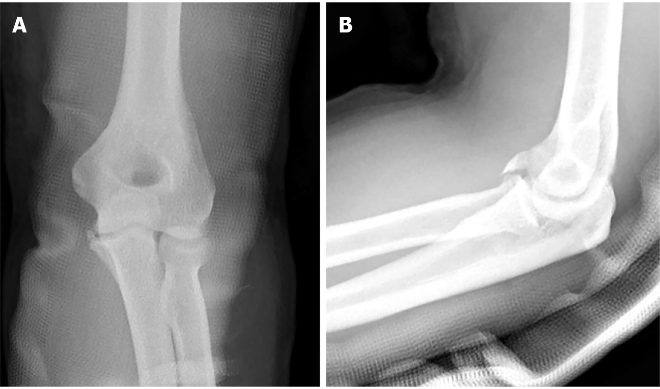
Figure 3 Injury films on presentation depicting a R&M type 2 coronoid fracture.
A and B: Anterior – posterior (A) and lateral (B) views.
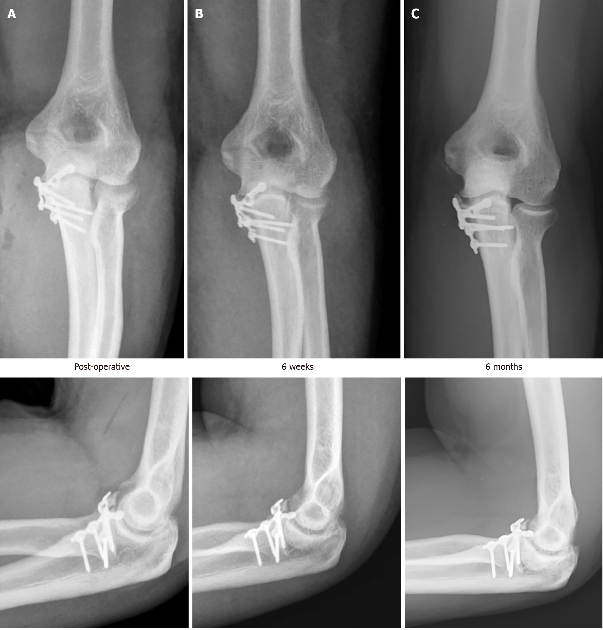
Figure 4 Post operative radiographs depicting progress.
A-C: Radiographs immediately post op (A), at 6 wk (B) and at 6 months (C).
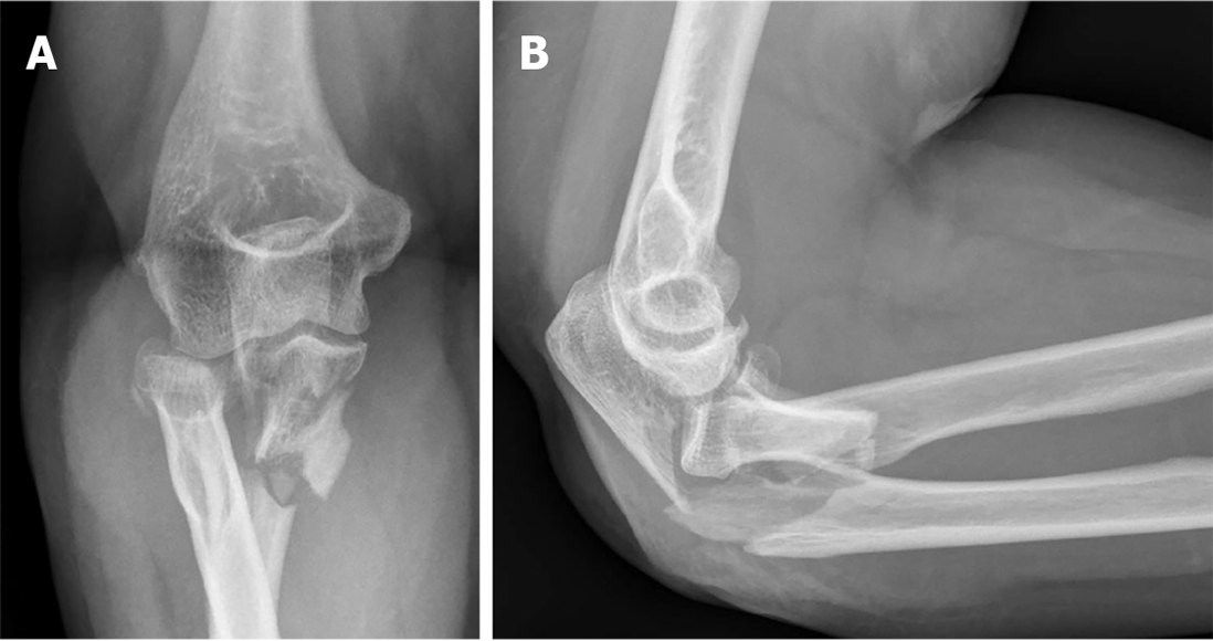
Figure 5 Injury films on presentation depicting a Monteggia variant type fracture.
A and B: Anterior – posterior (A) and lateral (B) views.
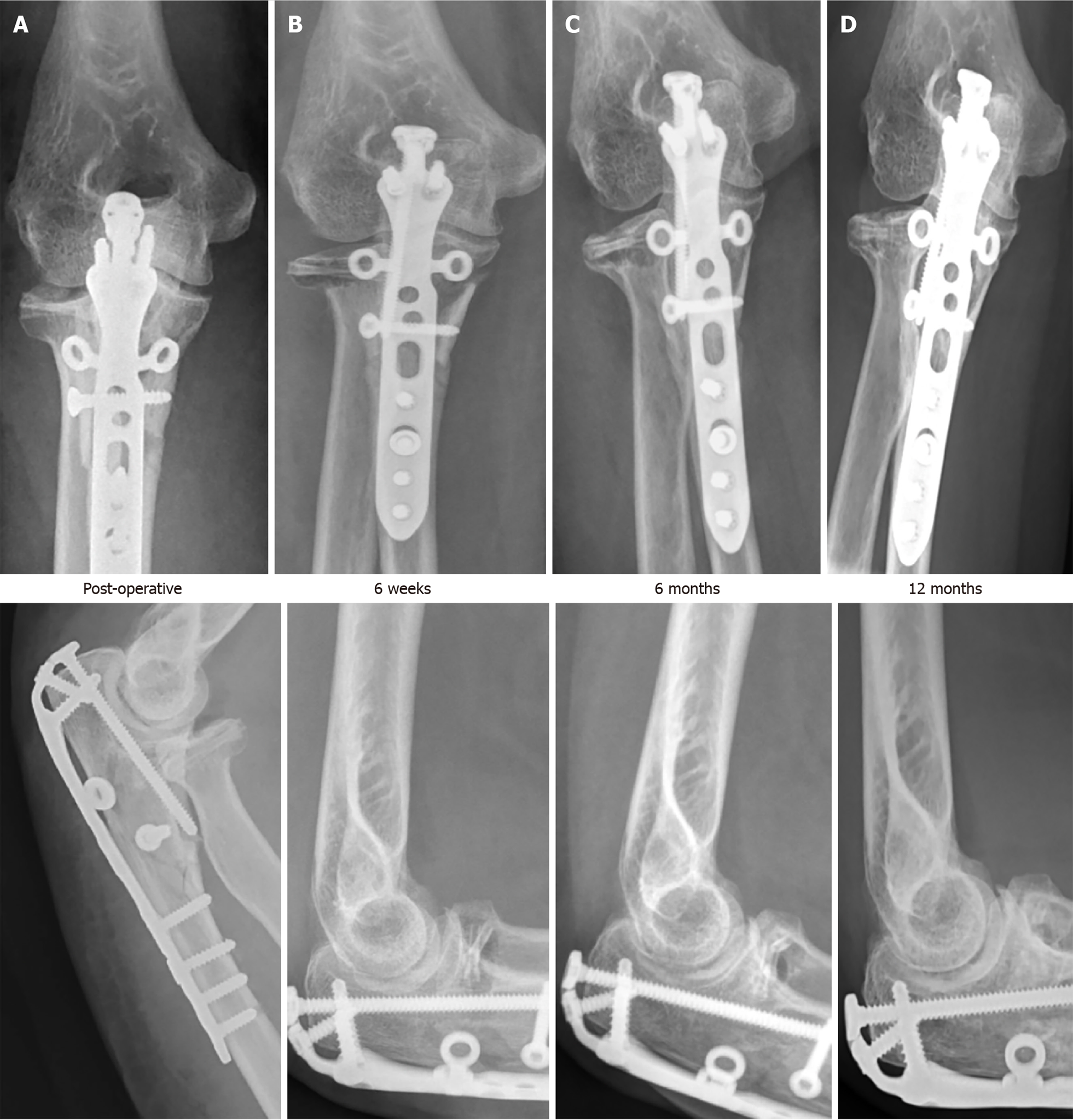
Figure 6 Post operative radiographs depicting progress.
A-D: Radiographs immediately post op (A), at 6 wk (B), 6 months (C) and 12 months (D).
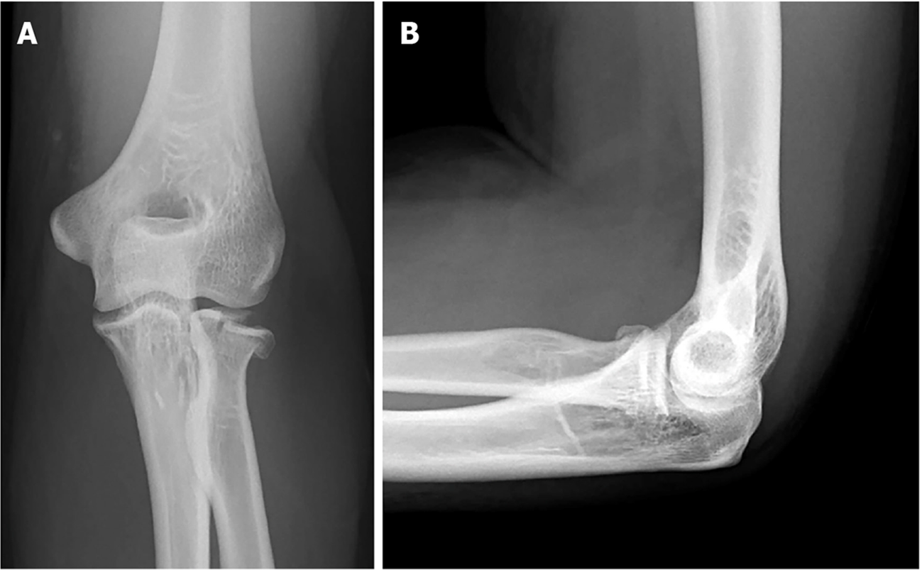
Figure 7 Injury films on presentation depicting a Mason type 2 radial head fracture.
A and B: Anterior – posterior (A) and lateral (B) views.
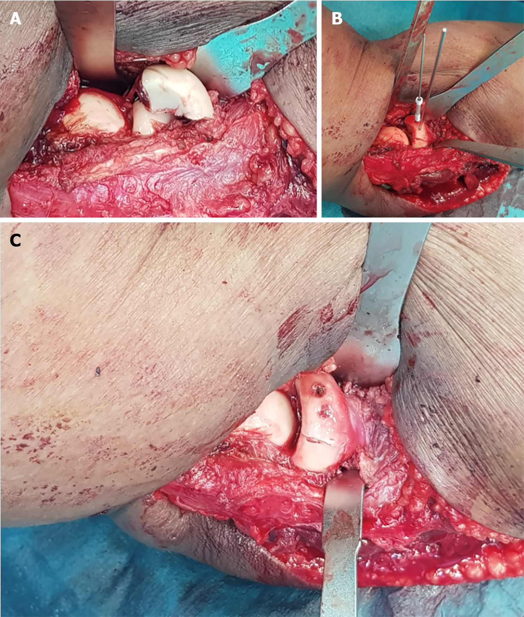
Figure 8 Intra-operative photos.
A: Initial fracture configuration; B and C: Reduction and fixation with cannulated magnesium screws (B) and finally the end result (C).
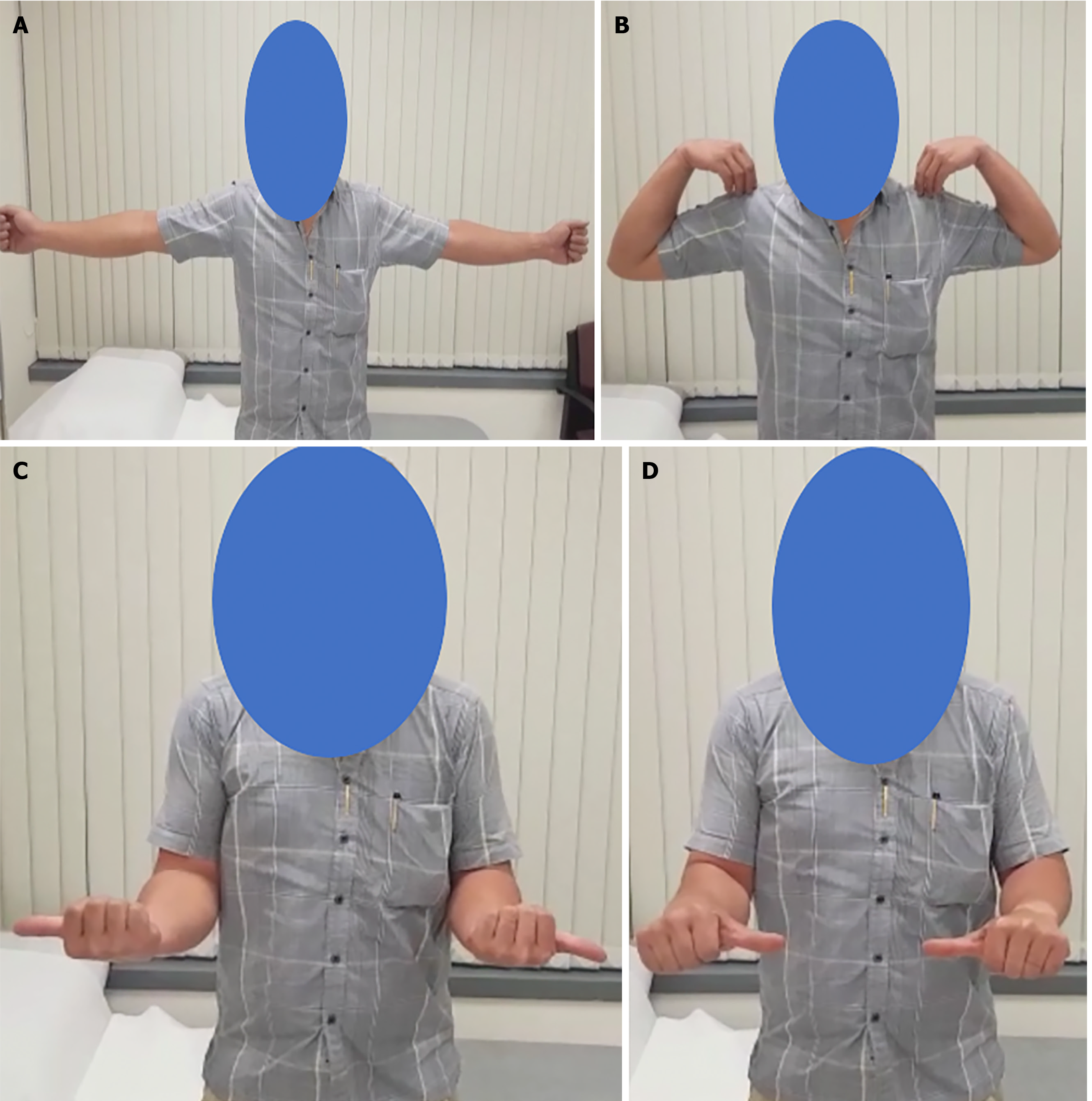
Figure 9 Post – operative range of motion at six wk post-operatively.
A: Elbow extension; B: Flexion; C: External rotation; D: Internal rotation.
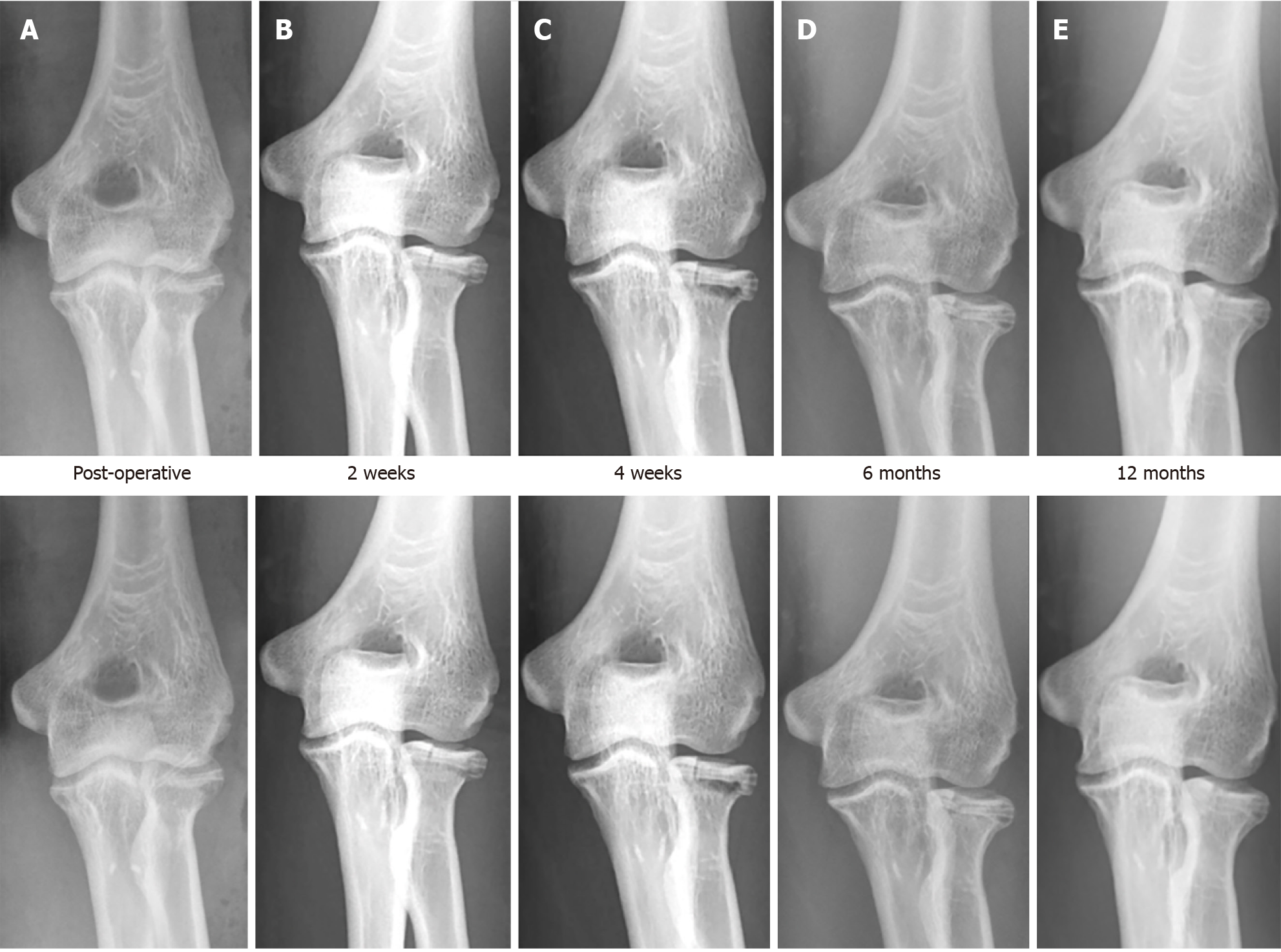
Figure 10 Post operative radiographs.
A-E: Radiographs immediately post op (A), at 2 weeks (B), 4 wk (C), 6 months (D) and 12 months (E).
Figure 11 Injury films on presentation depicting a terrible triad fracture.
A-D: Anterior posterior (A) and lateral (B) radiographs, saggital computer tomography cuts showing coronoid fracture (C) and radial head fracture (D).
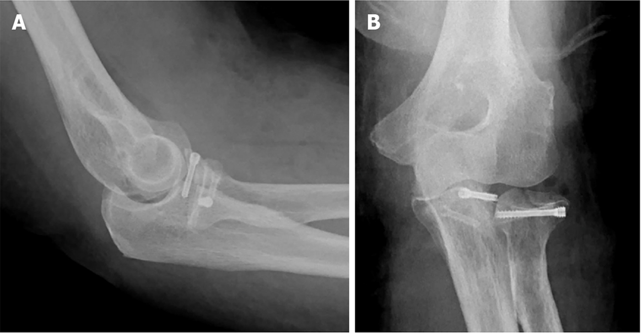
Figure 12 Immediate post-operative radiographs.
A and B: Anterior – posterior (A) and lateral (B) views.
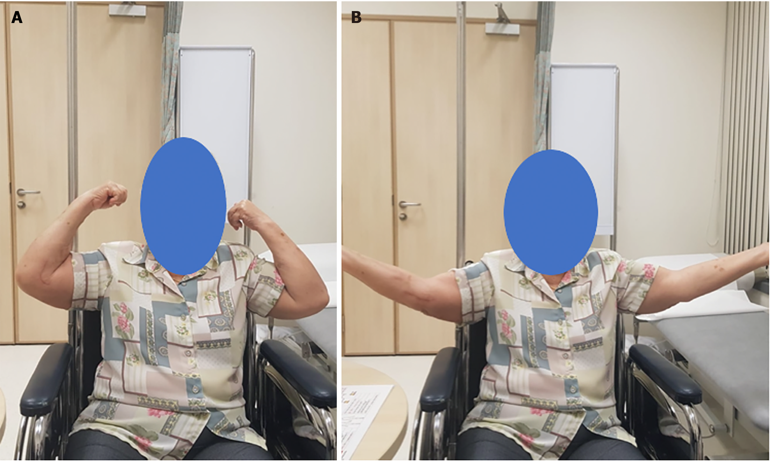
Figure 13 Post-operative range of motion at six months post operatively.
A and B: Elbow flexion (A) and extension (B).
Figure 14 Post operative radiographs.
A-E: Radiographs immediately post op (A), at 4 weeks (B), 4 months (C), 6 months (D) and 12 months (E).
- Citation: Fang C, Premchand AXR, Park DH, Toon DH. Peri-articular elbow fracture fixations with magnesium implants and a review of current literature: A case series. World J Orthop 2024; 15(3): 215-229
- URL: https://www.wjgnet.com/2218-5836/full/v15/i3/215.htm
- DOI: https://dx.doi.org/10.5312/wjo.v15.i3.215


