Copyright
©The Author(s) 2024.
World J Orthop. Dec 18, 2024; 15(12): 1175-1182
Published online Dec 18, 2024. doi: 10.5312/wjo.v15.i12.1175
Published online Dec 18, 2024. doi: 10.5312/wjo.v15.i12.1175
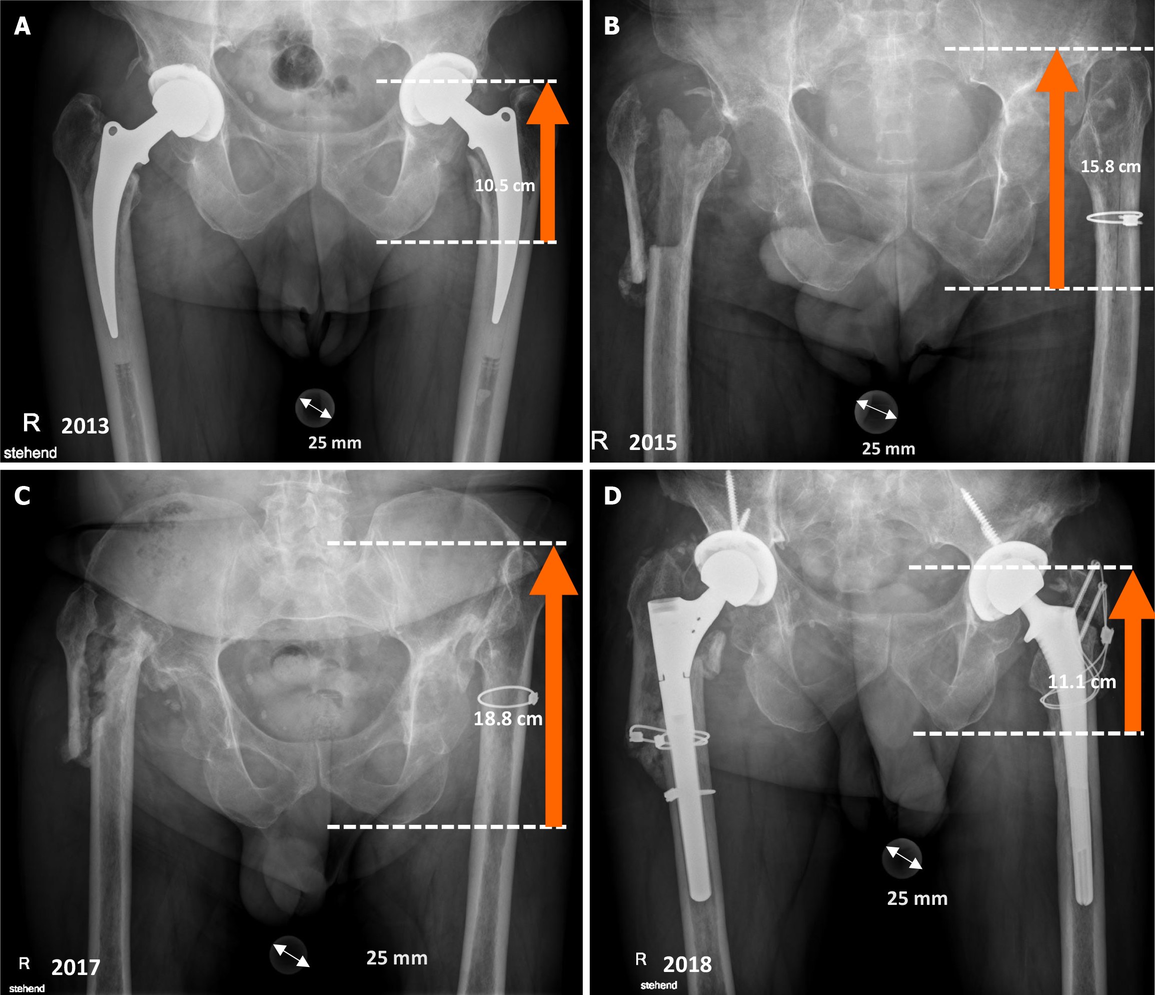
Figure 1 A 58-year-old patient experienced poor functional outcomes following Girdlestone resection arthroplasty.
Weight bearing X-rays of the 58-year-old patient from 2013-2018. The dotted white lines and the orange arrows indicate the distance between the ischial tuberosity and the greater trochanter. A: After Girdlestone resection arthroplasty (GRA) in 2013. The baseline distance between the ischial tuberosity and greater trochanter was 10.5 cm; B: After GRA in 2015. The distance between the ischial tuberosity and greater trochanter increased to 15.8 cm; C: After GRA in 2017. The baseline distance between the ischial tuberosity and greater trochanter increased to 18.8 cm, indicating a leg shortening of 8.3 cm; D: After reimplantation of bilateral revision hip arthroplasty.
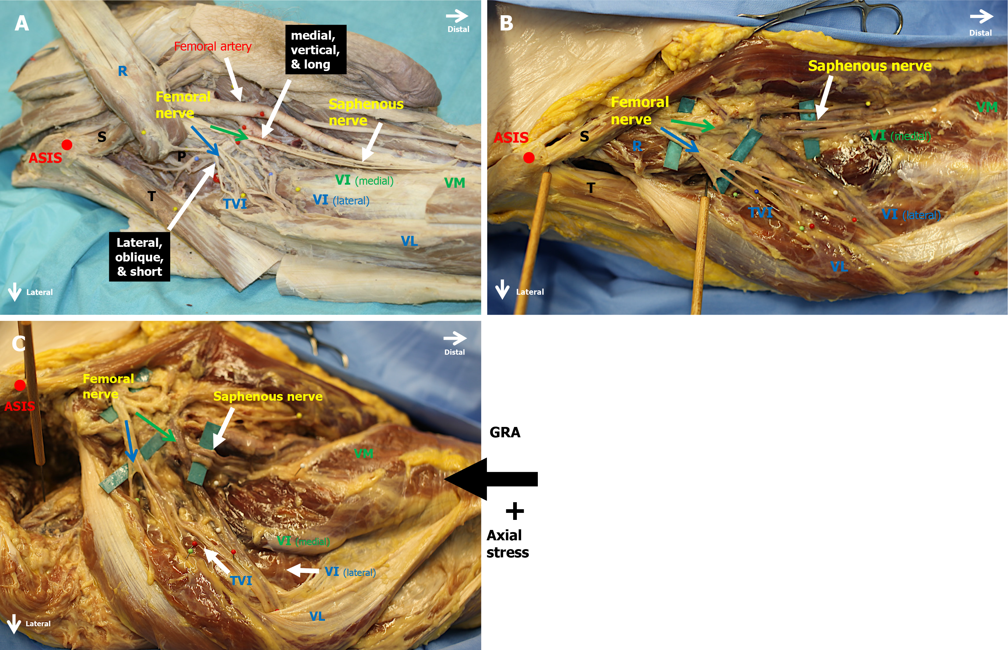
Figure 2 View of the right femoral thigh.
A: Antero-lateral view of the right femoral thigh. The anatomy of the femoral nerve with the medial vertically running division (green arrow) and the lateral short oblique division (blue arrow) running over the major psoas muscle, which is an obstacle for these nerve branches. The rectus femoris muscle (R) was retracted proximally and medially for better vision; B: Antero-lateral view of the right femoral thigh demonstrating the medial (green arrow) and lateral (blue arrow) femoral nerve divisions and its entry points into the muscle before Girdlestone resection arthroplasty. The nerve branches were highlighted with pieces of green paper; C: Antero-lateral view of the right thigh with the medial (green arrow) and lateral (blue arrow) division of the femoral nerve after Girdlestone resection arthroplasty. The femoral nerve was highlighted with green paper. We performed a Girdlestone resection arthroplasty through a direct anterior approach to the hip joint and applied axial stress to the calcaneus (thick black arrow). We measured the distance from the anterior superior iliac spine (red point) to the nerve entry points into the muscle. ASIS: Anterior superior iliac spine; GRA: Girdlestone resection arthroplasty; TVI: Tensor vastus intermedius; VI (lateral): Vastus intermedius lateral part; VI (medial): Medial area of the vastus intermedius; VM: Vastus medialis.
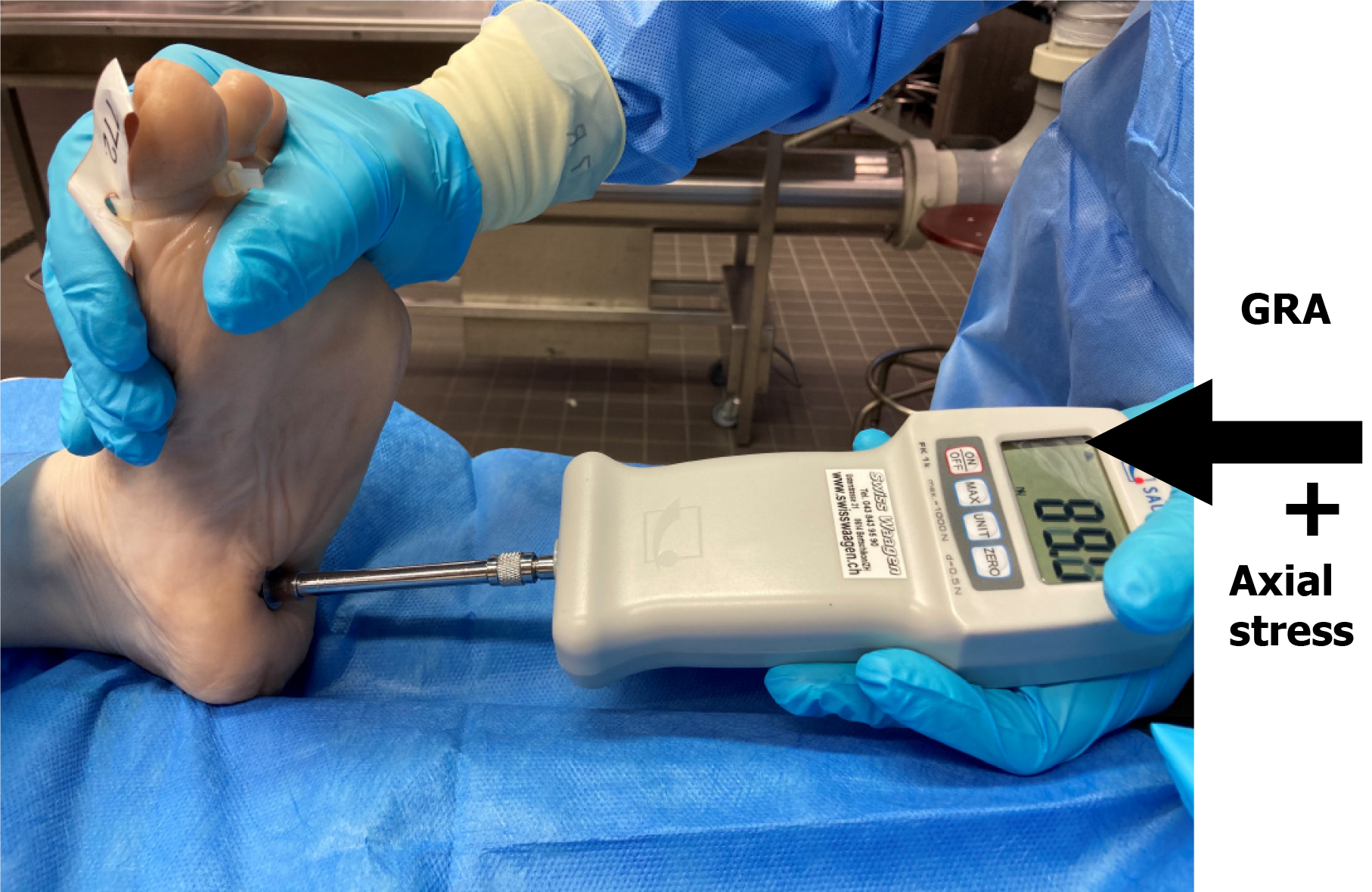
Figure 3 Demonstration of an axial force application to a left calcaneus with a digital force gauge.
Measurements were made in Newtons (Sauter FK, Swiss Waagen DC GmbH). GRA: Girdlestone resection arthroplasty.
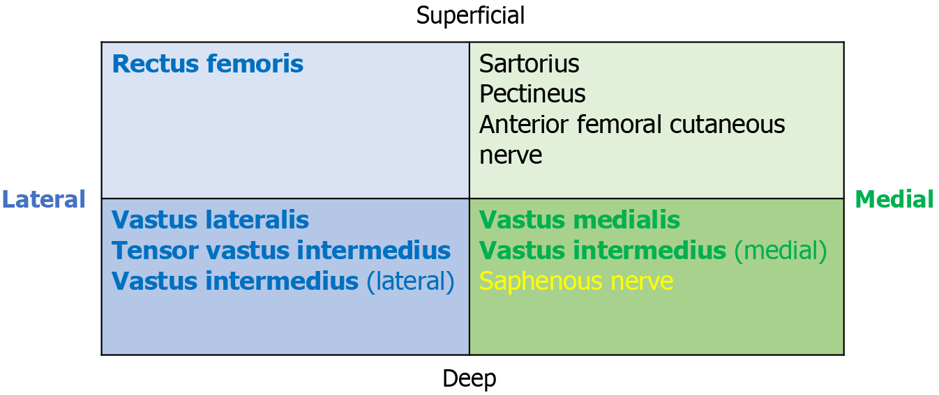
Figure 4 Schematic cross-section of the femoral nerve.
It consists of medial and lateral areas subdivided into superficial and deep portions. The rectus femoris, vastus lateralis, tensor vastus intermedius, and lateral area of the vastus intermedius are located in the lateral division (blue). The medial area of the vastus intermedius and the vastus medialis are located in the medial division (green). The anterior femoral cutaneous nerve and saphenous nerve are sensory nerve branches, and the sartorius and pectineus muscles are supplied by the superficial medial division.
- Citation: Spuehler D, Kuster L, Ullrich O, Grob K. Femoral nerve palsy following Girdlestone resection arthroplasty: An observational cadaveric study. World J Orthop 2024; 15(12): 1175-1182
- URL: https://www.wjgnet.com/2218-5836/full/v15/i12/1175.htm
- DOI: https://dx.doi.org/10.5312/wjo.v15.i12.1175


