Copyright
©The Author(s) 2024.
World J Orthop. Dec 18, 2024; 15(12): 1118-1123
Published online Dec 18, 2024. doi: 10.5312/wjo.v15.i12.1118
Published online Dec 18, 2024. doi: 10.5312/wjo.v15.i12.1118
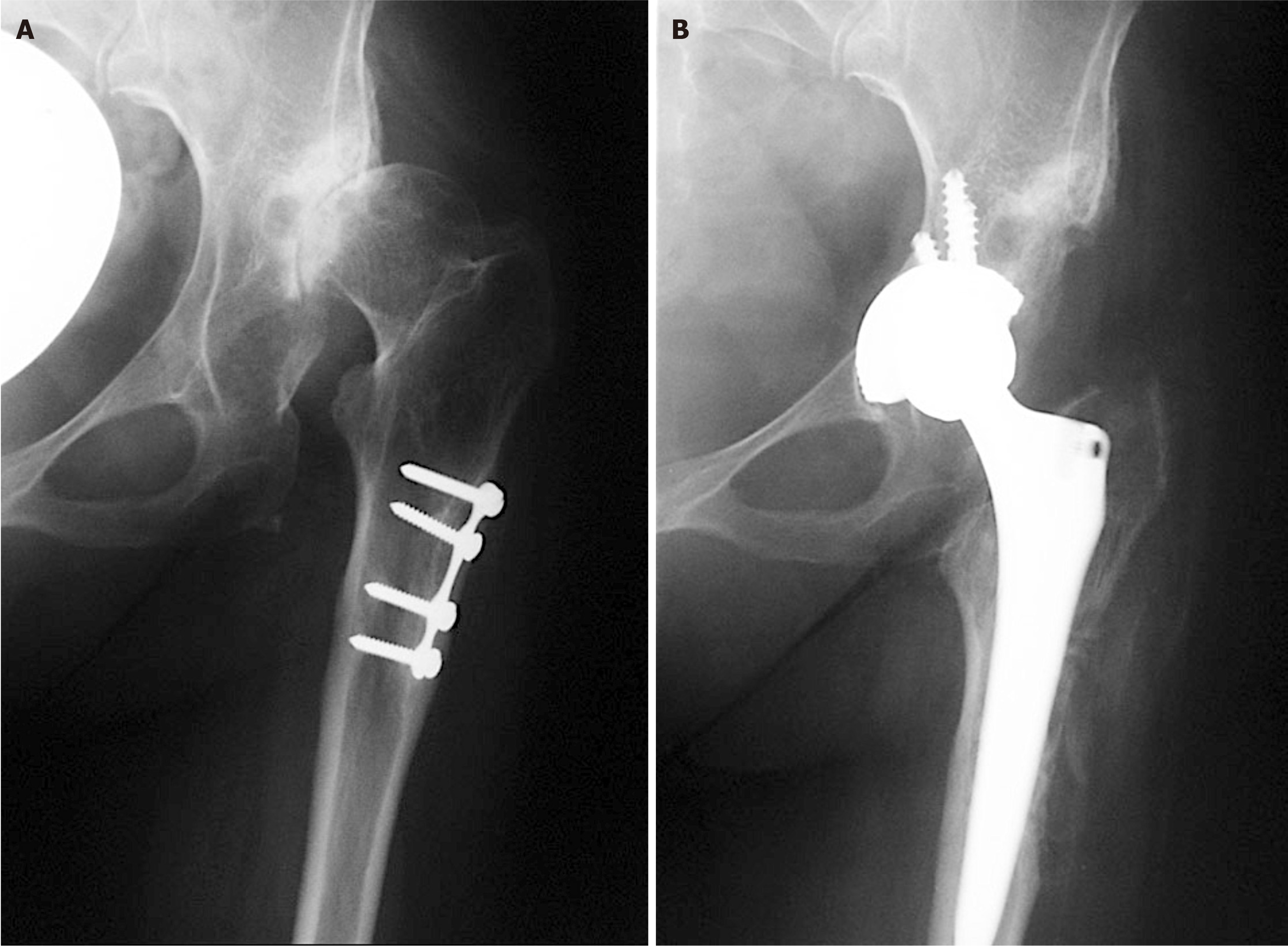
Figure 1 X-rays of left hip.
A: Preoperative X-ray of a patient with secondary osteoarthritis of the left hip due to dysplasia. Plate and screws in the proximal femur are left from the previous surgery performed in childhood; B: Postoperative X-ray with implanted uncemented acetabular cup and femoral stem. Acetabular cup is protruding beyond the Kohler’s line inside the pelvis and secured with 3 additional screws. Lesser trochanter is brought distally to the normal level so there is no leg length discrepancy postoperatively.
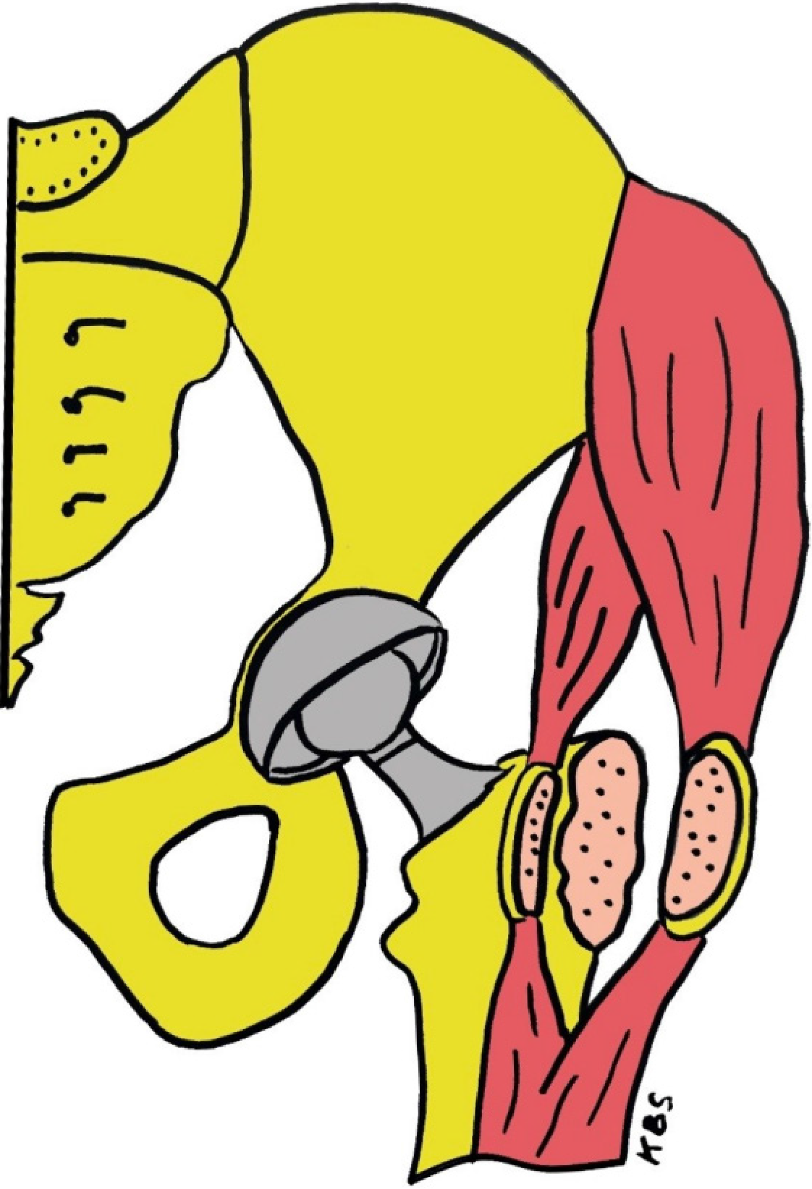
Figure 2 Drawing of our prefer method of treatment.
Acetabular cup is medialized (cotyloplasty) so that the dome of the cup is protruding beyond Kohler’s line inside the pelvis. On the femoral side anterior and posterior half of the continuous tendon is mobilized by a chisel. First the anterior part is divided with a thin layer of bone from the greater trochanter but remains attached to the continuous tendon of the gluteus medius and the vastus lateralis. And the same is done with the posterior half of the continuous tendon of the gluteus medius and the vastus lateralis which are detached with the chisel leaving a bone flake of at least 2 mm to 3 mm thickness attached to tendons. With that procedure continuity of abductor muscles is preserved and femur can be shortened without affecting the muscles. KBS: Critical region for definable segment defects.
- Citation: Barbaric Starcevic K, Bicanic G, Bicanic L. Specific approach to total hip arthroplasty in patients with childhood hip disorders sequelae. World J Orthop 2024; 15(12): 1118-1123
- URL: https://www.wjgnet.com/2218-5836/full/v15/i12/1118.htm
- DOI: https://dx.doi.org/10.5312/wjo.v15.i12.1118


