Copyright
©The Author(s) 2024.
World J Orthop. Nov 18, 2024; 15(11): 1101-1108
Published online Nov 18, 2024. doi: 10.5312/wjo.v15.i11.1101
Published online Nov 18, 2024. doi: 10.5312/wjo.v15.i11.1101
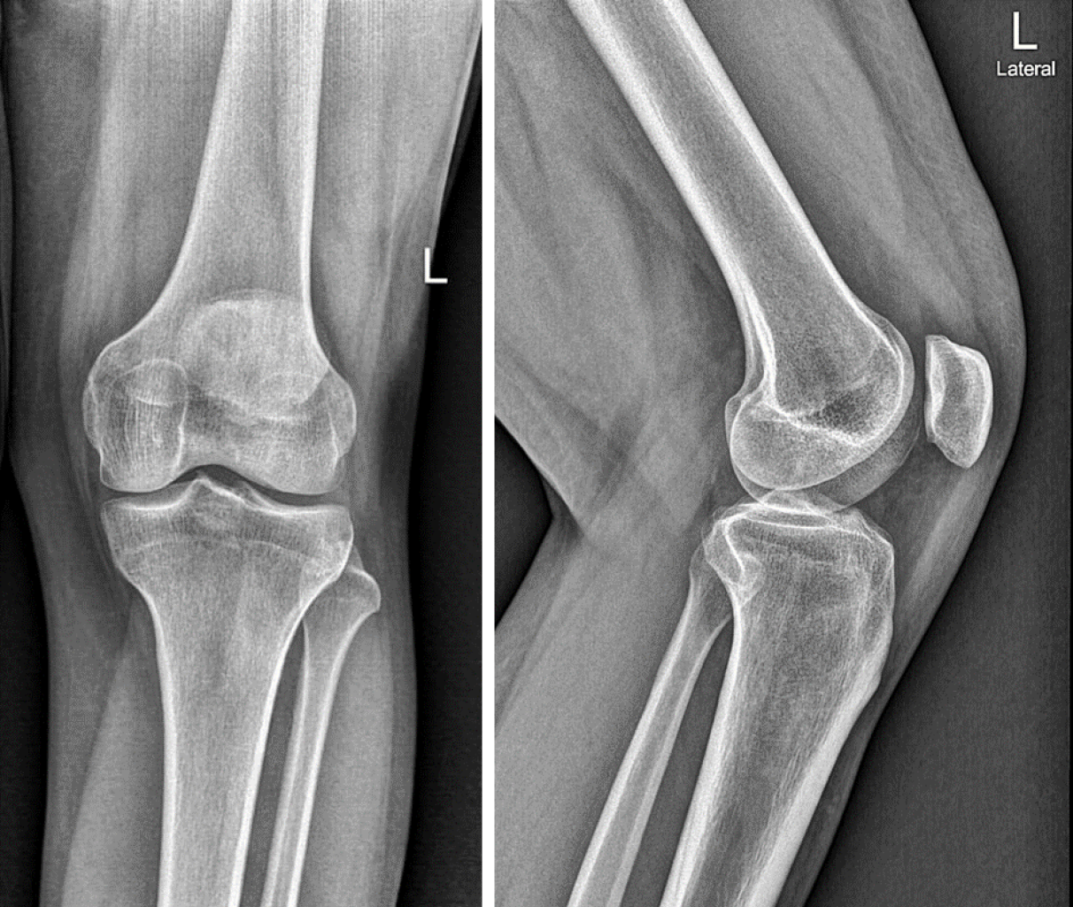
Figure 1 Plain radiography of the left knee revealed no abnormalities.
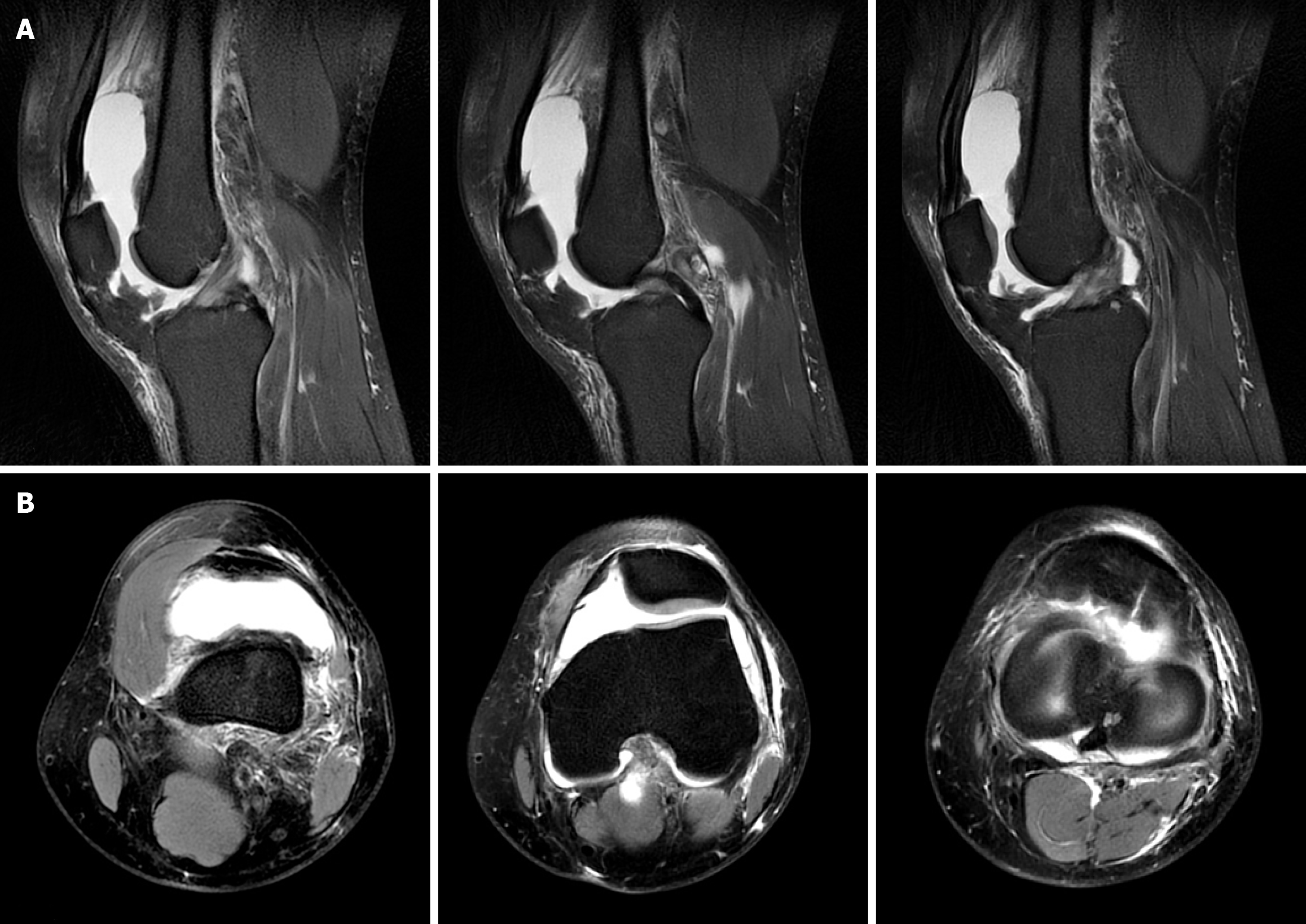
Figure 2 Preoperative magnetic resonance imaging revealed a large knee effusion in combination with synovitis.
A: Sagittal T2-weighted view; B: Axial T2-weighted view.
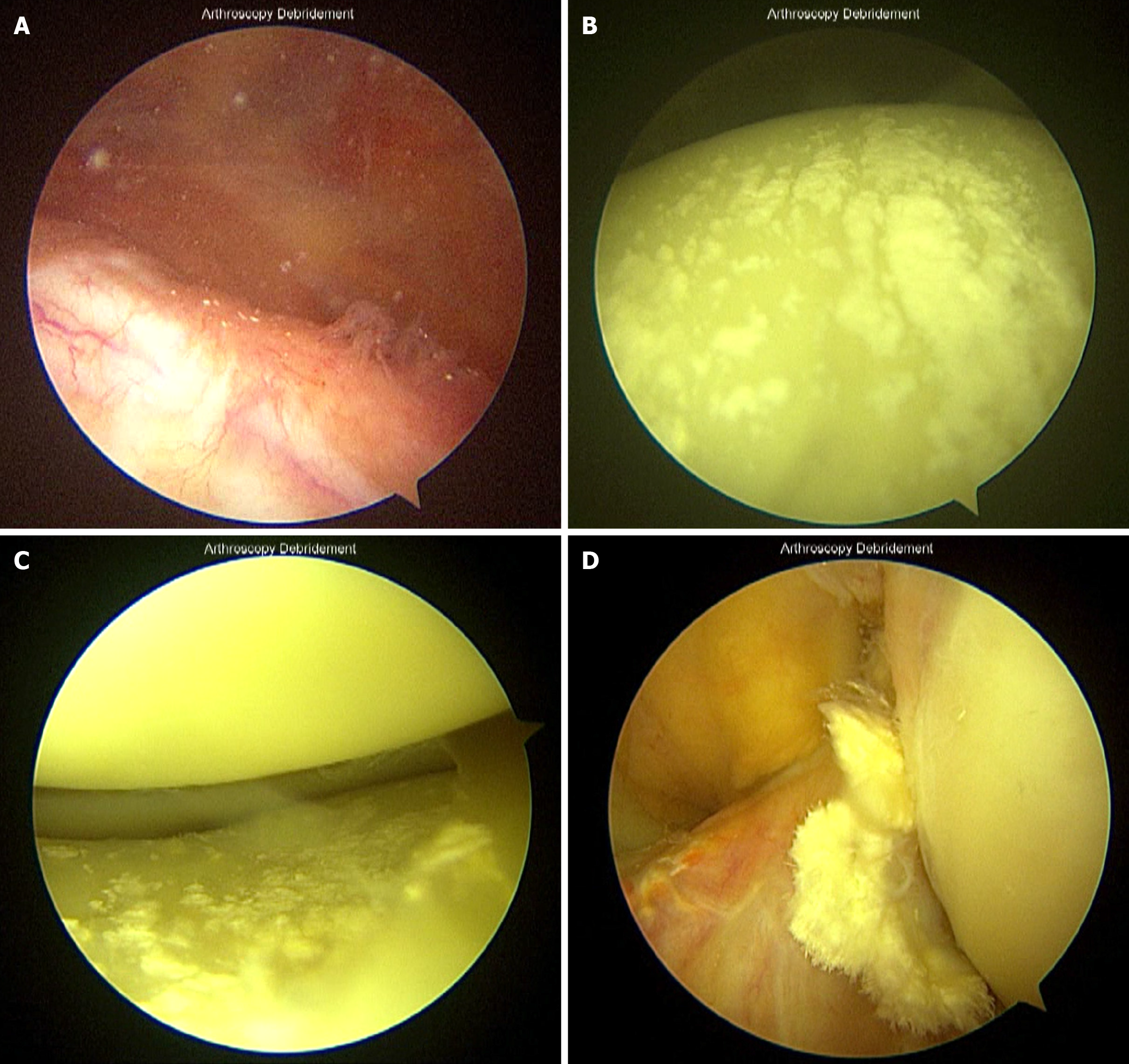
Figure 3 Diagnostic arthroscopy findings of chronic knee gouty arthritis.
A: Floating tophi were observed after the insertion of the viewing portal; B: Deposition of tophi on the cartilage surface of the femoral condyle; C: Deposition of tophi on the cartilage surface of the tibial plateau; D: Deposition of tophi on the anterior cruciate ligament surface.
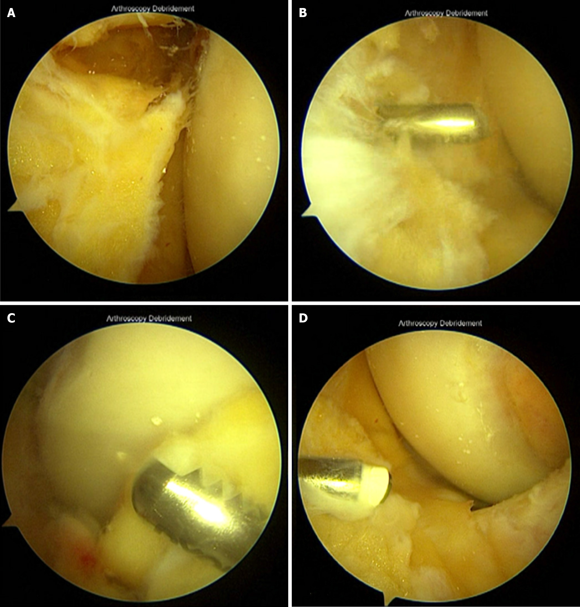
Figure 4 Arthroscopic synovectomy for synovial hyperplasia in chronic knee gouty arthritis.
A: Excessive synovial tissue was observed; B: Partial synovectomy was performed through the anteromedial portal; C: Partial synovectomy was also performed through the anterolateral portal; D: Radiofrequency was applied to the synovial surface after the thorough partial synovectomy.
- Citation: Utoyo GA, Calvin C. Arthroscopic synovectomy for synovial hyperplasia in chronic knee gouty arthritis: A case report. World J Orthop 2024; 15(11): 1101-1108
- URL: https://www.wjgnet.com/2218-5836/full/v15/i11/1101.htm
- DOI: https://dx.doi.org/10.5312/wjo.v15.i11.1101


