Copyright
©The Author(s) 2024.
World J Orthop. Nov 18, 2024; 15(11): 1088-1094
Published online Nov 18, 2024. doi: 10.5312/wjo.v15.i11.1088
Published online Nov 18, 2024. doi: 10.5312/wjo.v15.i11.1088
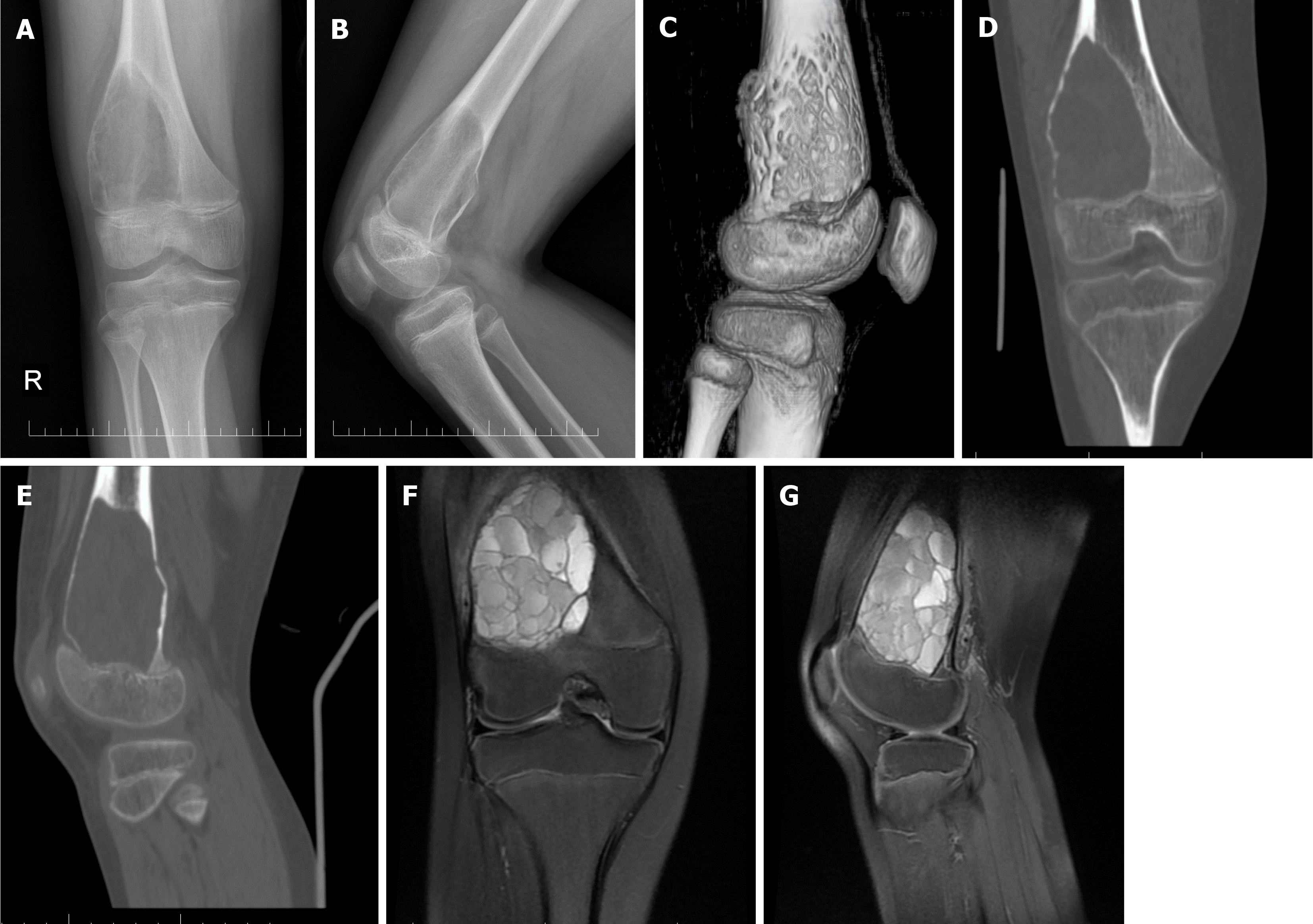
Figure 1 Preoperative images.
A: Anterior-posterior X-ray image of the right knee; B: Lateral X-ray view of the right knee; C: Computed tomography (CT) 3-dimensionalreconstruction of the right knee; D: Sagittal plane CT image of the right knee; E: Coronal plane CT of the right knee; F: Sagittal plane magnetic resonance imaging image of the right knee; G: Coronal plane CT image of the right knee.
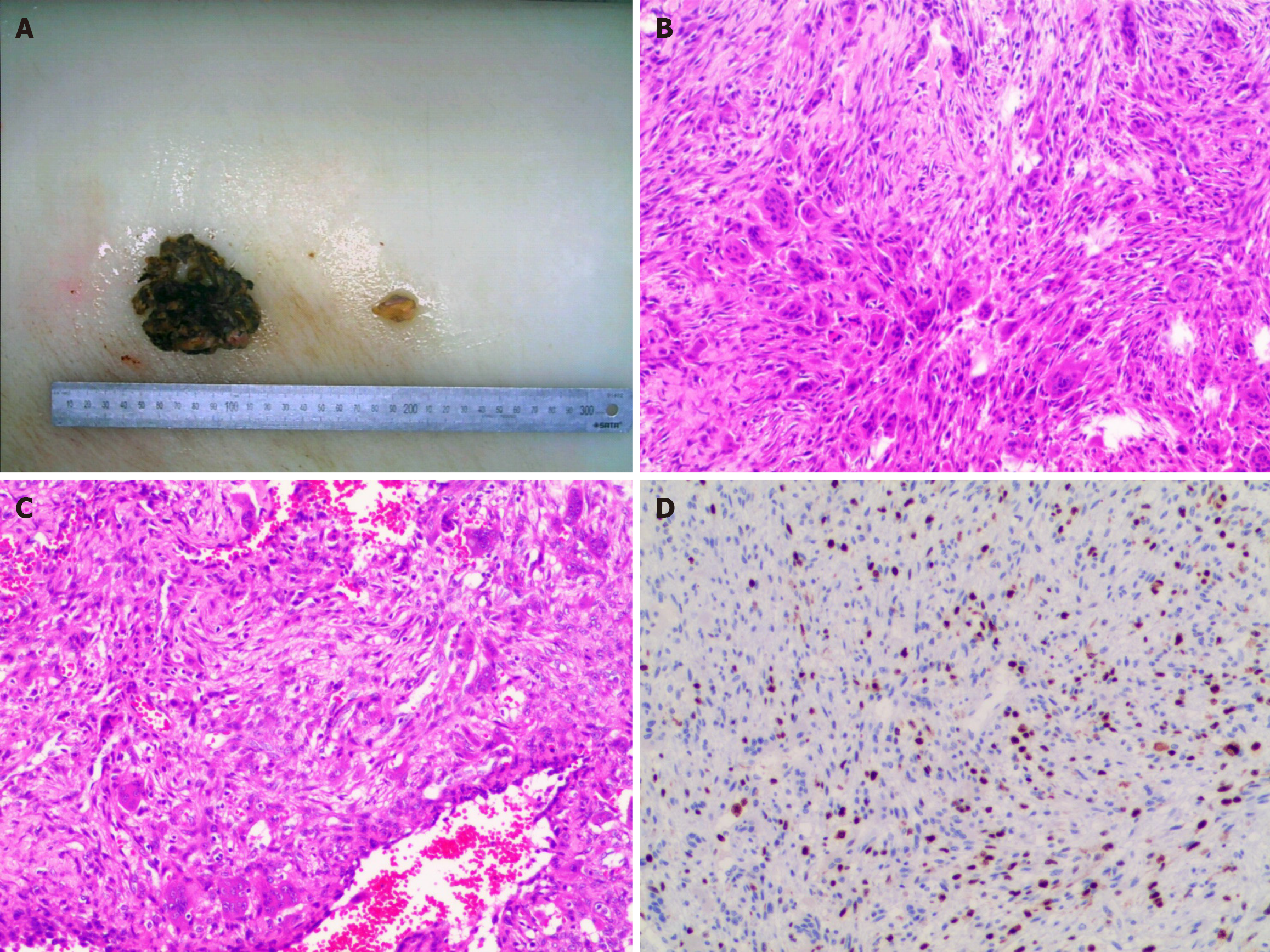
Figure 2 Pathological photos.
A: Appearance of the removed lesion; B-D: Microscopic view of the stained lesion.
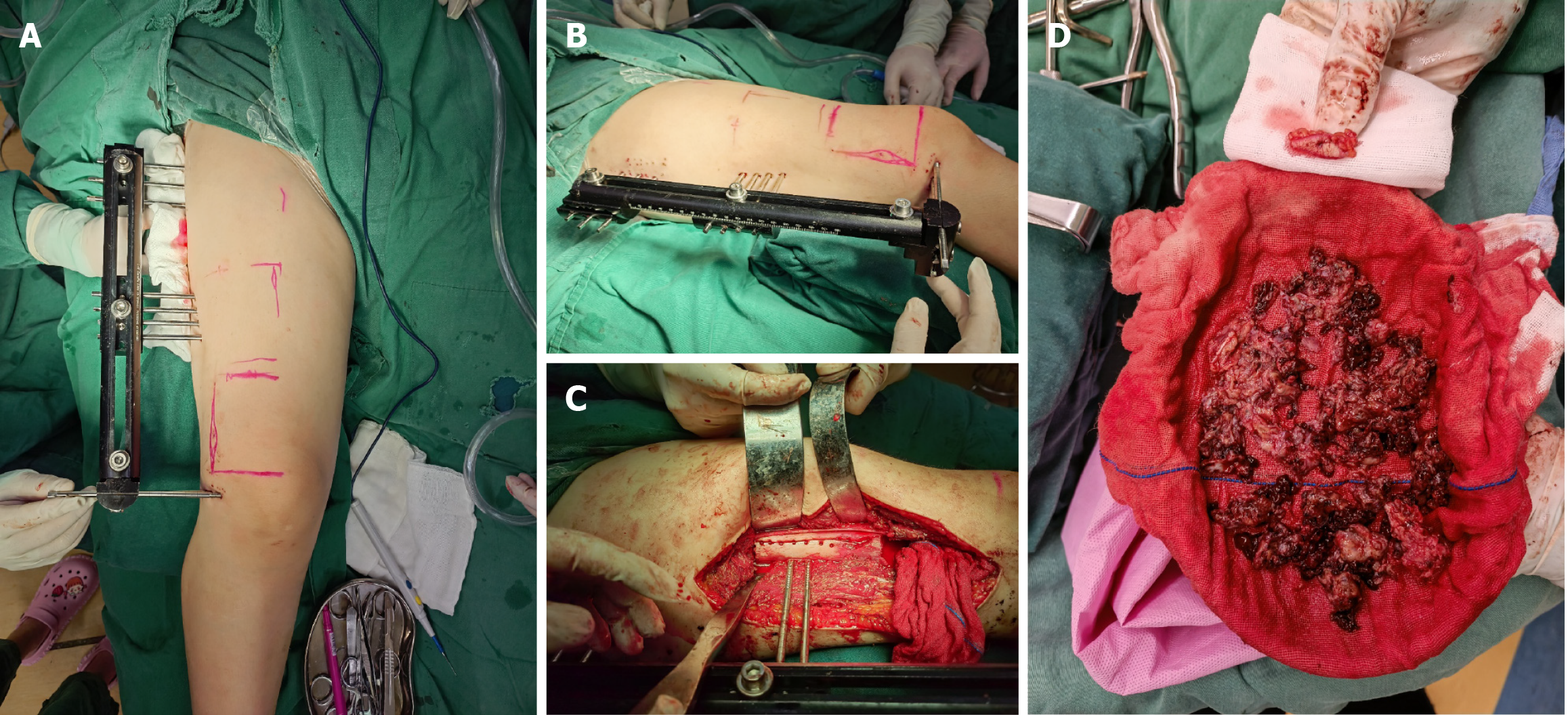
Figure 3 Intraoperative pictures.
A and B: Pictures after the external fixation frame; C: Picture of resection of tumor; D: The picture of extracted tumor tissue.
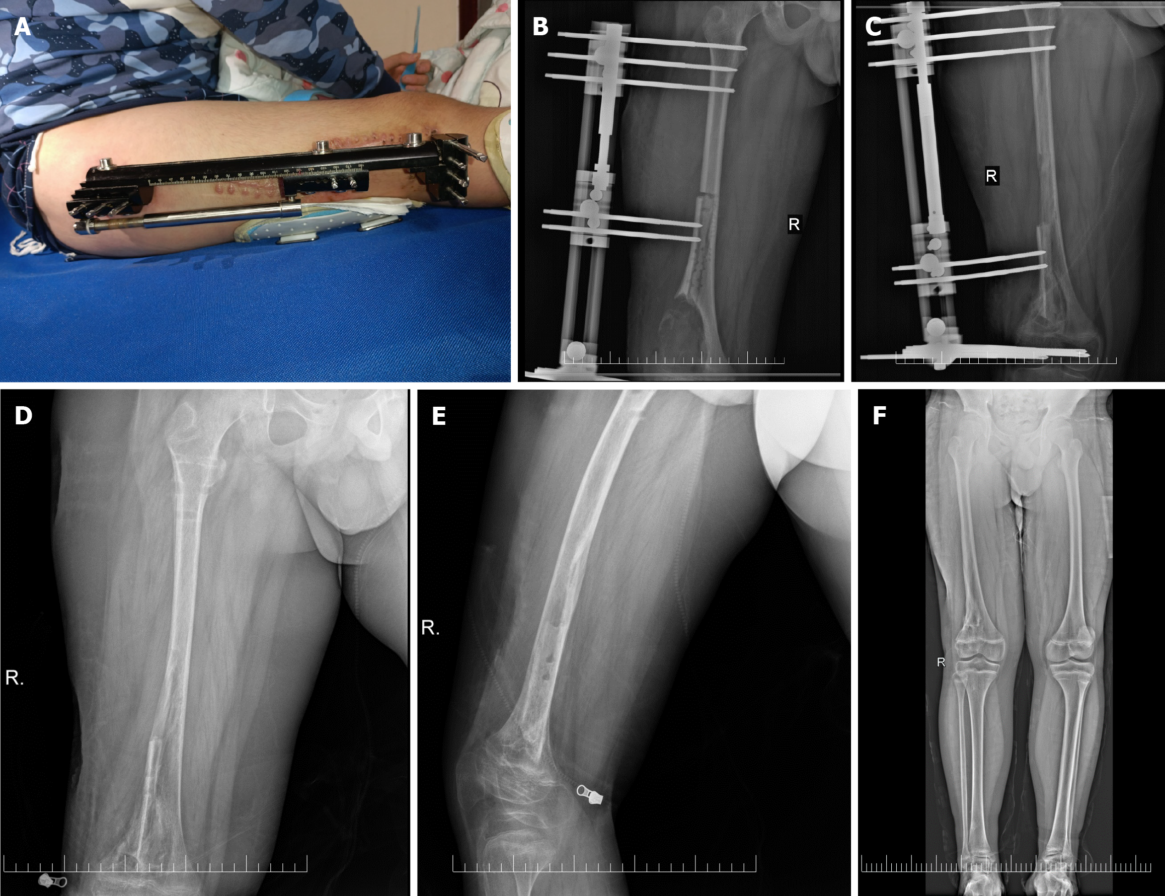
Figure 4 Postoperative follow-up photos.
A: Photo at review; B: Anterior-posterior X-ray image of the right knee four days after surgery; C: Anterior-posterior X-ray image of the right knee five months after surgery; D: Anterior-posterior X-ray image of the right knee after remove the external fixator; E: Lateral X-ray image of the right knee after remove the external fixator; F: Anterior-posterior X-ray image of the lower limbs.
- Citation: Long XY, Sun F, Wang T, Li P, Tian Z, Wu XW. Ilizarov technique for treatment of a giant aneurysmal bone cyst at the distal femur: A case report. World J Orthop 2024; 15(11): 1088-1094
- URL: https://www.wjgnet.com/2218-5836/full/v15/i11/1088.htm
- DOI: https://dx.doi.org/10.5312/wjo.v15.i11.1088


