Copyright
©The Author(s) 2024.
World J Orthop. Nov 18, 2024; 15(11): 1047-1055
Published online Nov 18, 2024. doi: 10.5312/wjo.v15.i11.1047
Published online Nov 18, 2024. doi: 10.5312/wjo.v15.i11.1047
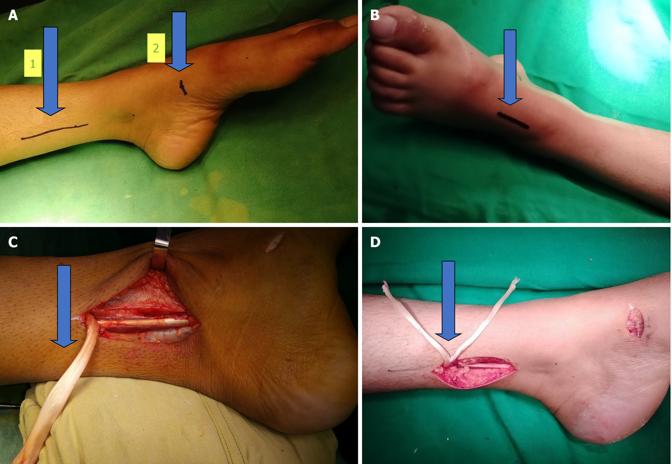
Figure 1 Photos of the tendon.
A: The arrow number 1 points towards the first incision which measures 5-7 cm. It is made along the palpable posteromedial border of the distal tibia. It ends about 2-3 cm proximal to the medial malleolus. The arrow number 2 indicates the second incision of 2 cm which is made over the navicular tuberosity (image date December 23, 2020); B: The third incision measures approximately 4 cm in length and is made over the dorsal aspect of the ankle to expose the tibialis anterior, extensor hallucis longus, extensor digitorum longus and peroneus tertius tendons (image date December 23, 2020); C: This clinical photograph shows the tibialis posterior tendon which has been disinserted from the navicular tuberosity and retracted proximally through the aforementioned first incision (image date December 23, 2020); D: This photograph shows the harvested tibialis posterior tendon which has been split into two slips (image date December 23, 2020).
- Citation: Saaiq M. Presentation and management outcome of foot drop with tibialis posterior tendon transfer. World J Orthop 2024; 15(11): 1047-1055
- URL: https://www.wjgnet.com/2218-5836/full/v15/i11/1047.htm
- DOI: https://dx.doi.org/10.5312/wjo.v15.i11.1047


