Copyright
©The Author(s) 2024.
World J Orthop. Nov 18, 2024; 15(11): 1036-1046
Published online Nov 18, 2024. doi: 10.5312/wjo.v15.i11.1036
Published online Nov 18, 2024. doi: 10.5312/wjo.v15.i11.1036
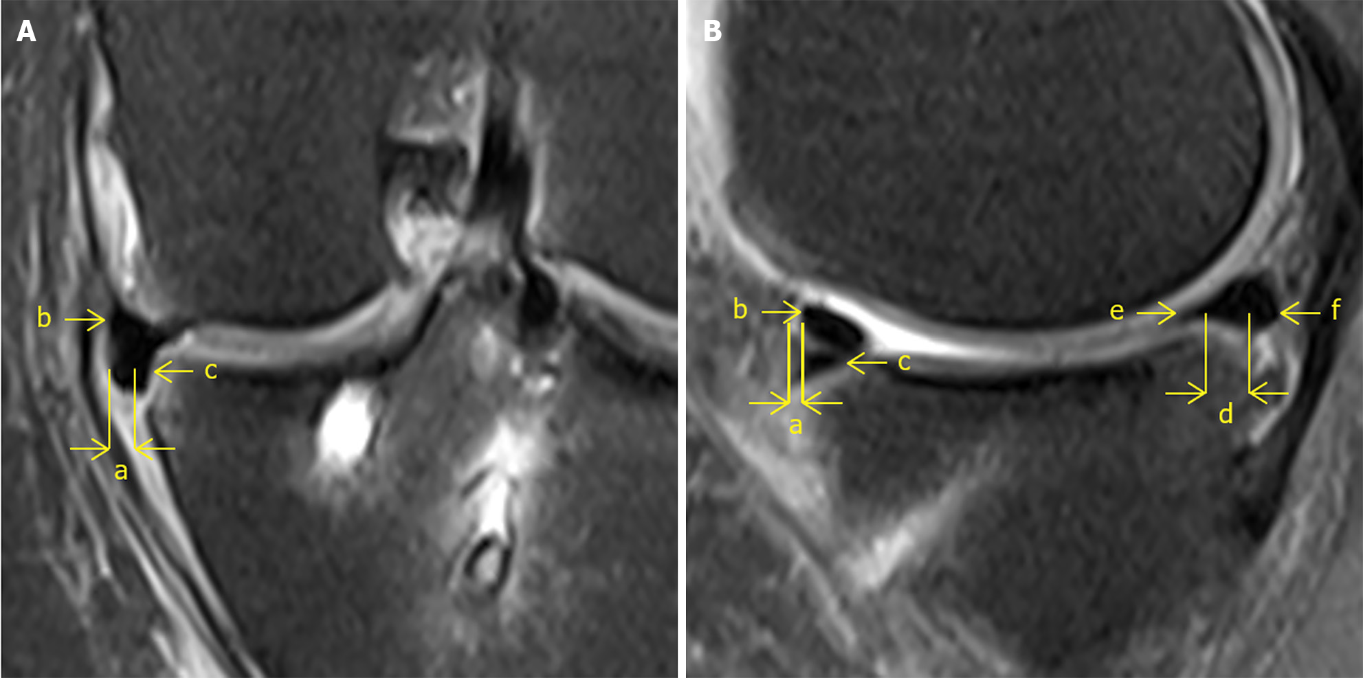
Figure 1 The absolute value of the degree of graft extrusion, measured as the horizontal distance from the most peripheral aspect of the graft to the edge of the tibia measured on the slice that the medial joint space is the narrowest on coronal and sagittal knee magnetic resonance imaging.
A: Medial extrusion. a: The absolute value of medial extrusion; b: Meniscus remnant; c: Tendon graft; B: Anterior and posterior extrusiona. a: The absolute value of anterior extrusion; b: Tendon graft; c: Meniscus remnant; d: The absolute value of posterior extrusion; e: Tendon graft; f: Meniscus remnant.
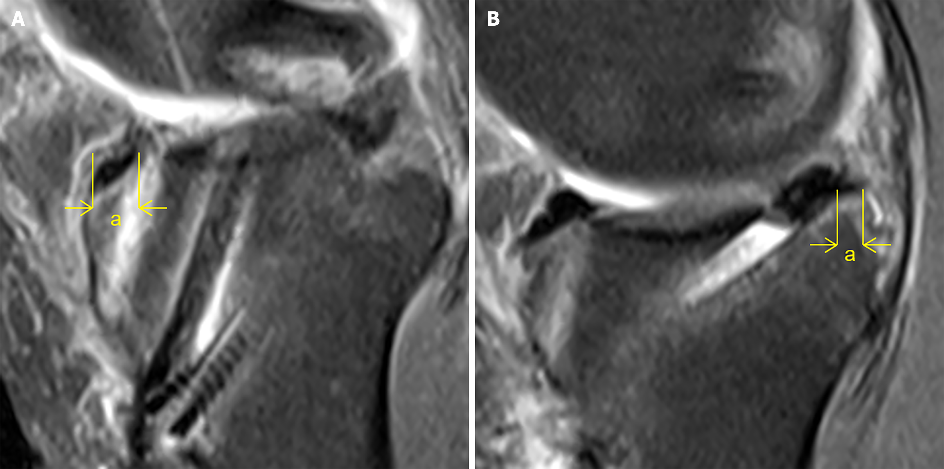
Figure 2 Tunnel edge distance, measured as the horizontal distance from the intraarticular tunnel entrance to the edge of the tibia measured on the slice where the tunnel entrance is the widest on sagittal knee magnetic resonance imaging.
A: Anterior tunnel edge distance (indicated by the letter “a”); B: Posterior tunnel edge distance (indicated by the letter “a”).
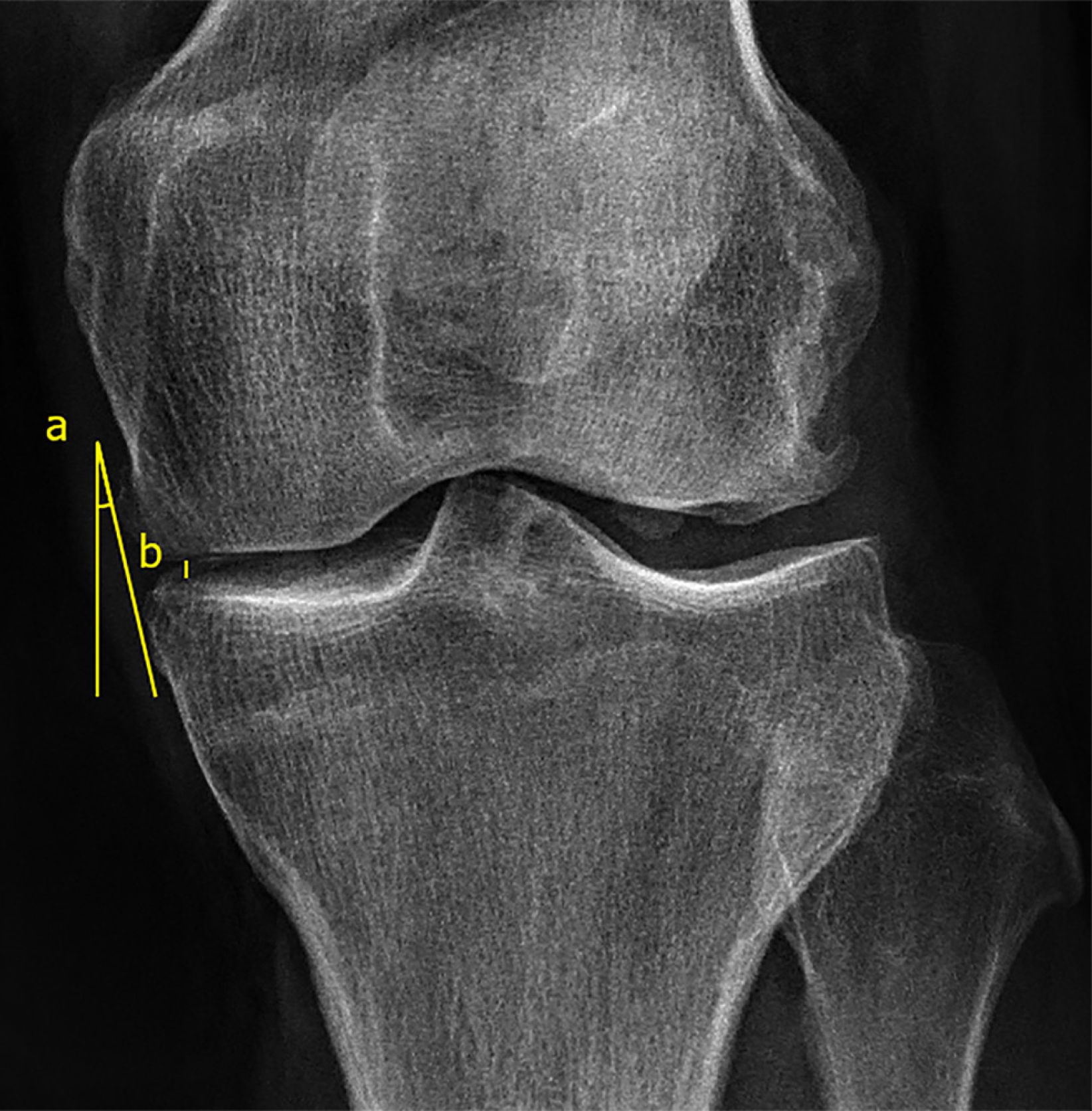
Figure 3 Medial edge incline angle and medial joint space measured on anteroposterior knee radiograph.
a: Medial edge incline angle, measured as the degree of the incline angle formed by the common tangent to the medial edge of the femur and tibia and the vertical line; b: Medial joint space, measured as the length of the vertical line segment from the medial edge of the tibia plateau to the femoral condyle.
- Citation: Zhu TW, Xiang XX, Li CH, Li RX, Zhang N. Predictive factors for coronal and sagittal graft extrusion length after using tendon autograft for medial meniscus reconstruction. World J Orthop 2024; 15(11): 1036-1046
- URL: https://www.wjgnet.com/2218-5836/full/v15/i11/1036.htm
- DOI: https://dx.doi.org/10.5312/wjo.v15.i11.1036


