Copyright
©The Author(s) 2024.
World J Orthop. Oct 18, 2024; 15(10): 981-990
Published online Oct 18, 2024. doi: 10.5312/wjo.v15.i10.981
Published online Oct 18, 2024. doi: 10.5312/wjo.v15.i10.981
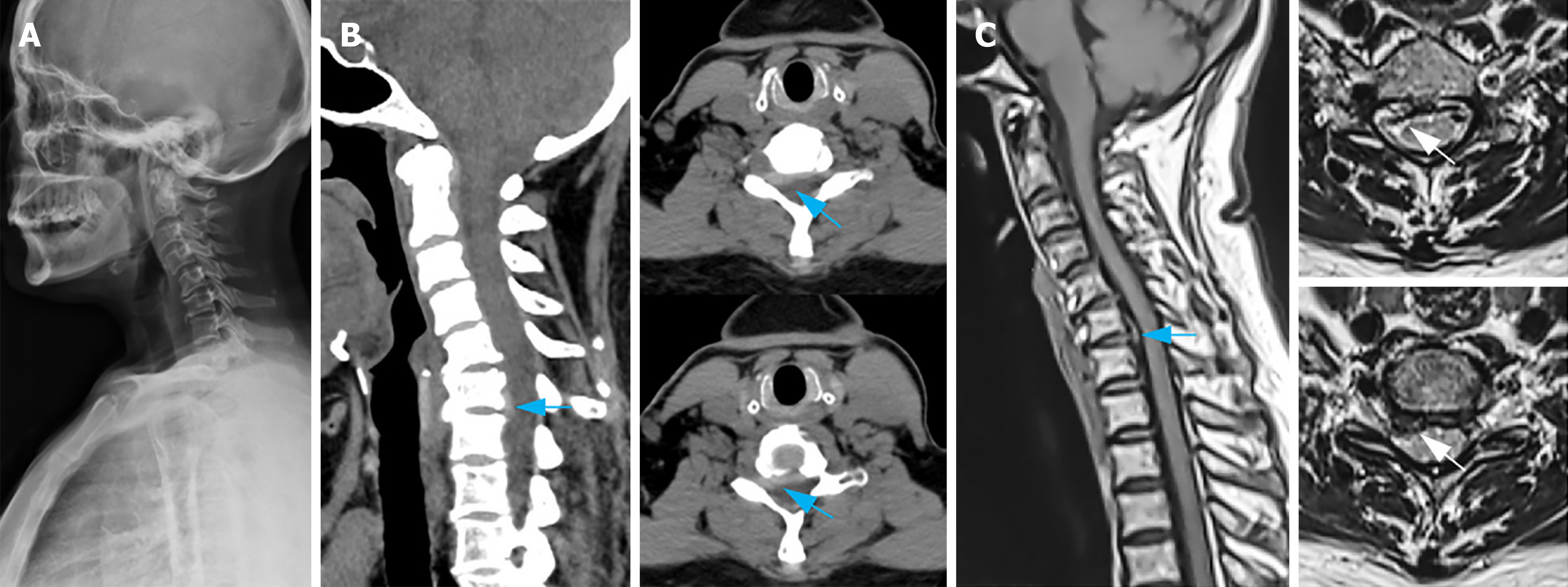
Figure 1 Preoperative imaging examinations.
A: A lateral plain film of the cervical vertebra depicts cervical vertebra's reversed arch, narrowed C6/7 space, and patchy calcification of the nuchal ligament; B: Cervical vertebra computerized tomography sagittal reconstruction and plain scan demonstrate an intervertebral disc prolapse and upward displacement in the C6/7 space, along with right nerve root canal stenosis (indicated by the blue arrow); C: Sagittal magnetic resonance imaging and plain scan of cervical vertebra present herniation and upward displacement of the intervertebral disc in the C6/7 space (indicated by the blue arrow), discontinuous intervertebral disc signal, and compression of the right C7 nerve root (indicated by the white arrow).
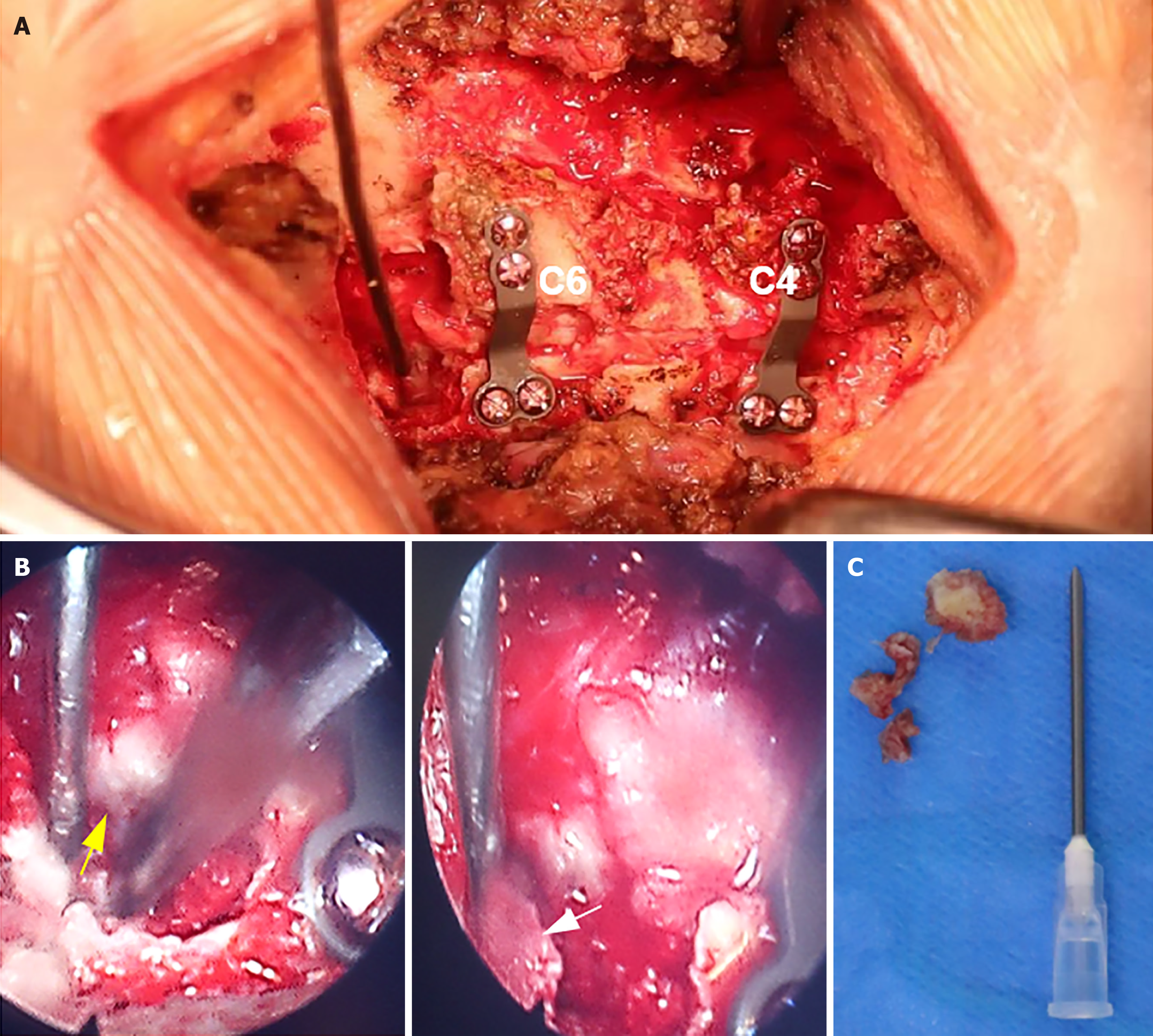
Figure 2 Intraoperative image.
A: The vertebral laminae of C4 and C6 were elevated, facilitating decompression of the spinal cord, followed by placement of an internal fixation plate; B: The ventral nucleus pulposus was extracted from the right nerve root of C7 using foraminal mirror-assisted extraction technique. During this procedure, the dural membrane (indicated by the yellow arrow) and excised free nucleus pulposus tissue (indicated by the white arrow) were visually observed; C: Intraoperatively obtained free nucleus pulposus tissue from the ventral aspect of the C7 nerve root.
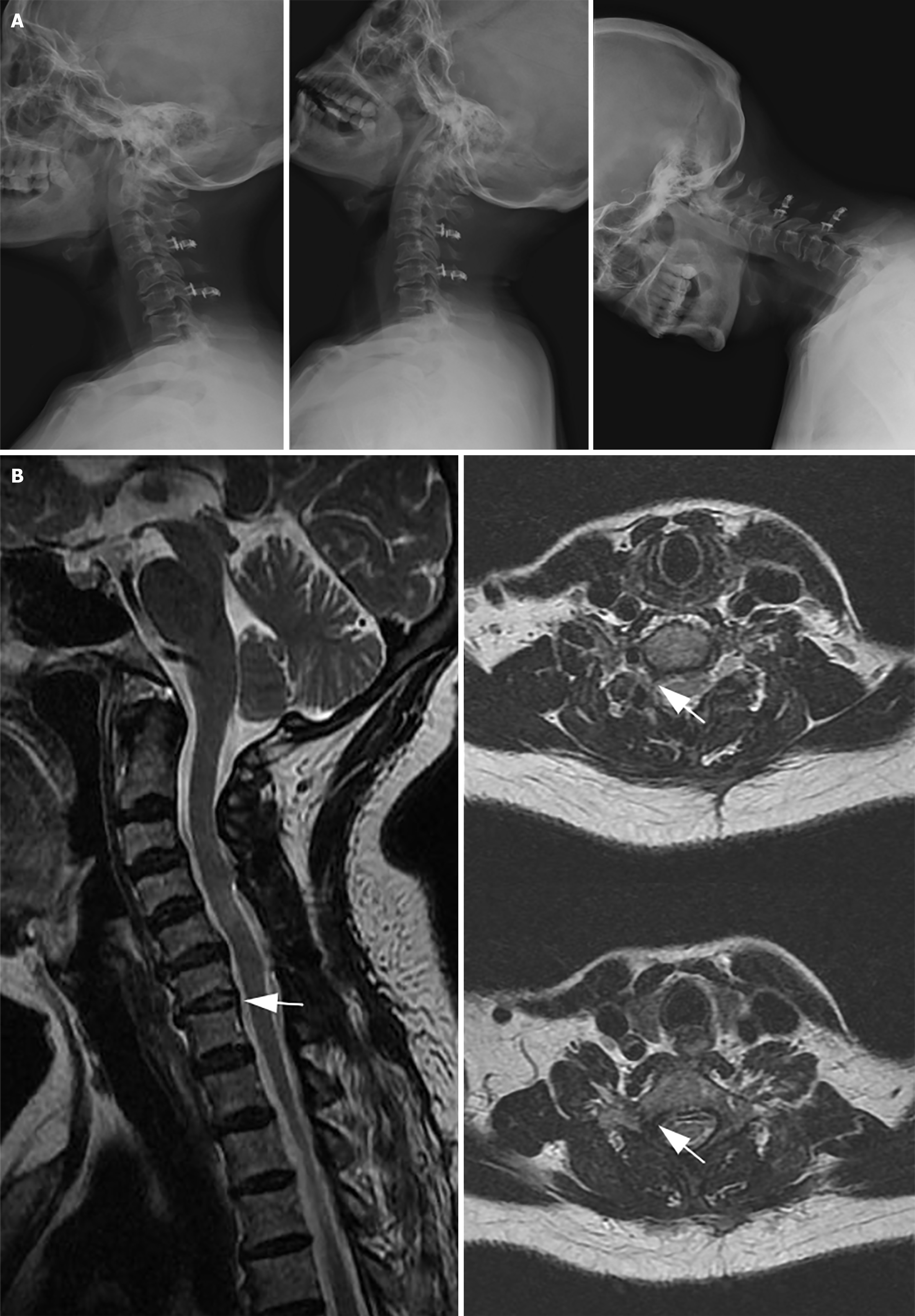
Figure 3 Outpatient follow-up 6 months post-surgery.
A: X-ray examination of the cervical spine in lateral and hyperextension flexion positions displays a significant improvement in the cervical spine's range of motion; B: Magnetic resonance imaging at 6 months post-surgery: The herniated nucleus pulposus tissue of C6/7 has been eliminated, and the right C7 nerve has been entirely decompressed, with no signal from the nucleus pulposus tissue (indicated by the white arrow).
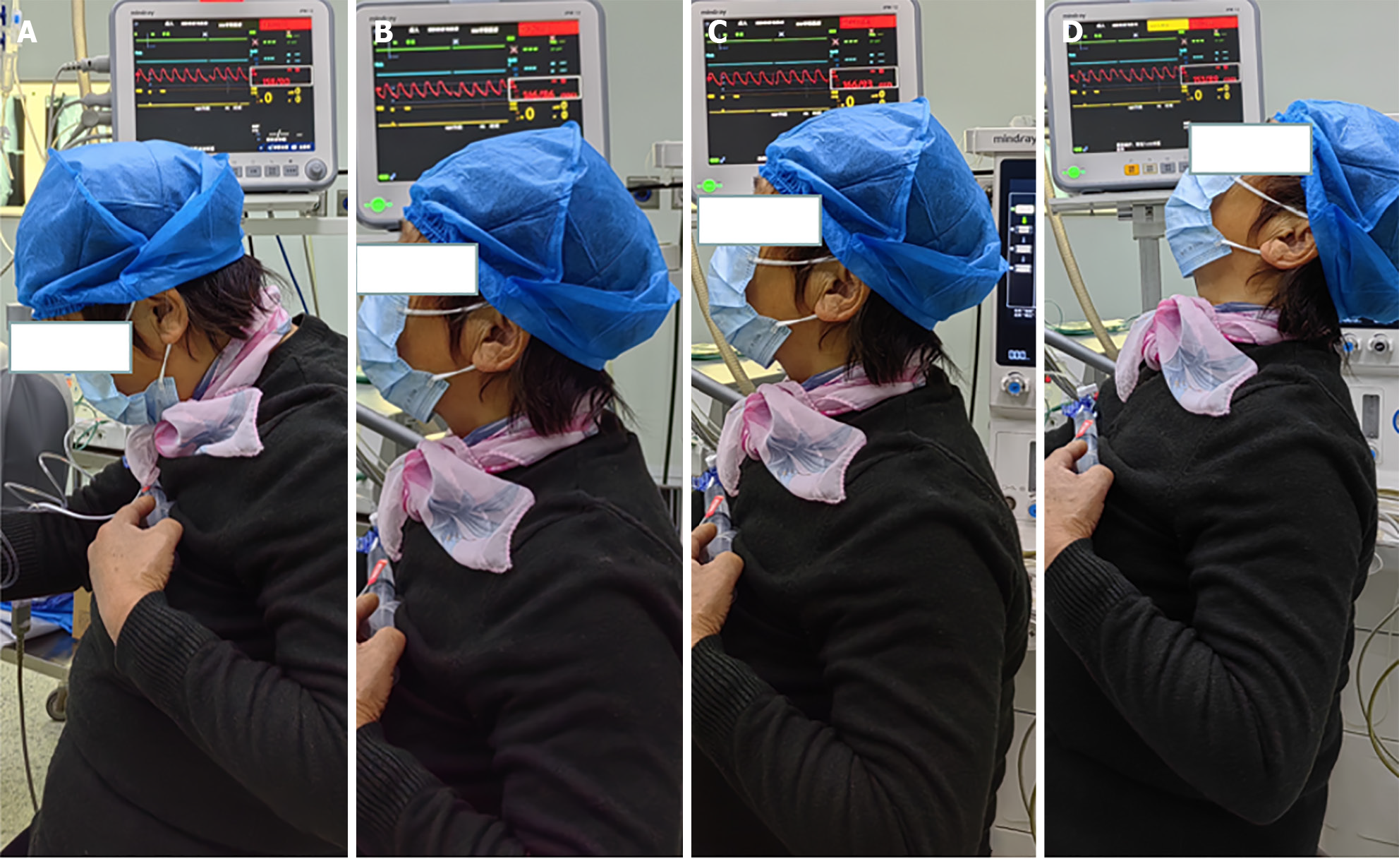
Figure 4 Six months post-surgery, ambulatory pulse blood pressure monitoring indicated systolic blood pressure fluctuations between 146–166 mmHg and diastolic blood pressure variations between 86–93 mmHg.
A: Cervical flexion, blood pressure at 158/90 mmHg; B: Neck in neutral position, blood pressure at 146/86 mmHg; C: Neck slightly extended: Blood pressure at 166/93 mmHg; D: Neck in complete extension, blood pressure at 153/89 mmHg.
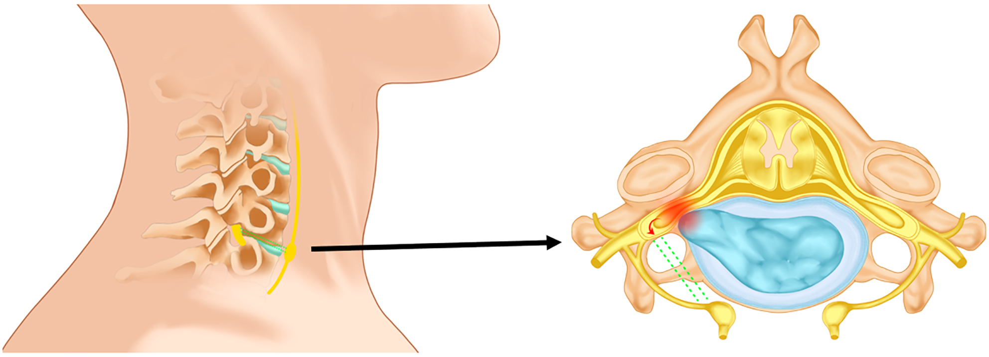
Figure 5 Overextension of the neck leads to further compression of the spinal ganglia by the nucleus pulposus tissue.
Following pressure stimulation, the spinal nerves directly excite the neck's sympathetic ganglia via bidirectional nerve fibers (represented by green dashed lines), thus activating the sympathetic nervous system and inducing a rapid blood pressure increase.
- Citation: Cui HC, Chang ZQ, Zhao SK. Atypical cervical spondylotic radiculopathy resulting in a hypertensive emergency during cervical extension: A case report and review of literature. World J Orthop 2024; 15(10): 981-990
- URL: https://www.wjgnet.com/2218-5836/full/v15/i10/981.htm
- DOI: https://dx.doi.org/10.5312/wjo.v15.i10.981


