Copyright
©The Author(s) 2024.
World J Orthop. Jan 18, 2024; 15(1): 94-100
Published online Jan 18, 2024. doi: 10.5312/wjo.v15.i1.94
Published online Jan 18, 2024. doi: 10.5312/wjo.v15.i1.94
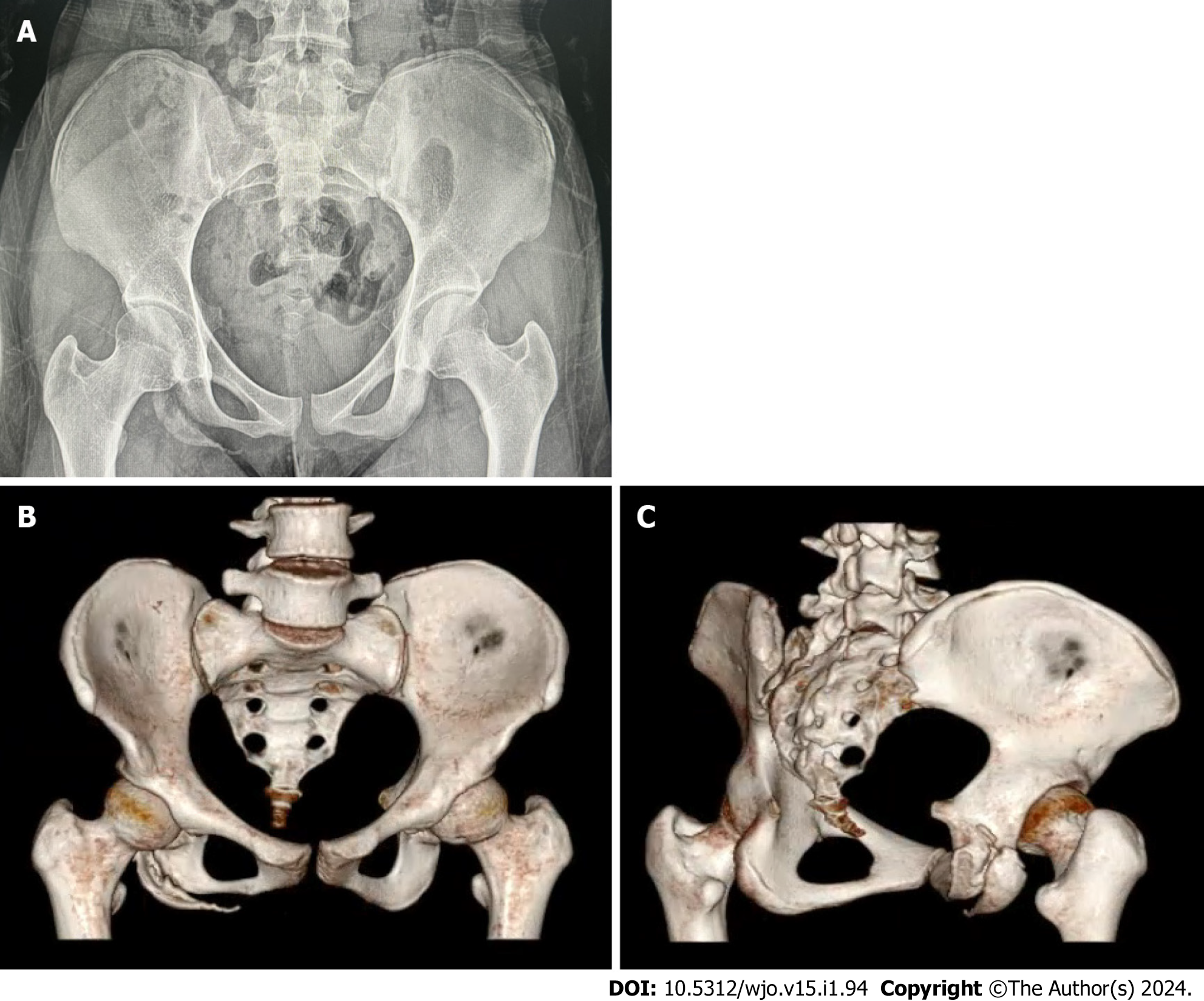
Figure 1 Imaging examinations photos.
A: Plain X-ray film of the pelvis, revealing an unhealed avulsion fracture of ischial tuberosity and partial ischial ramus. The fragment is displaced anterolaterally by approximately 2 cm; B and C: Computed tomography scan and 3D reconstruction of the pelvis exhibiting a fracture nonunion of the ischial tuberosity and partial ischial ramus, with the fragment measuring 6.4 cm × 1.3 cm × 2.9 cm in size.
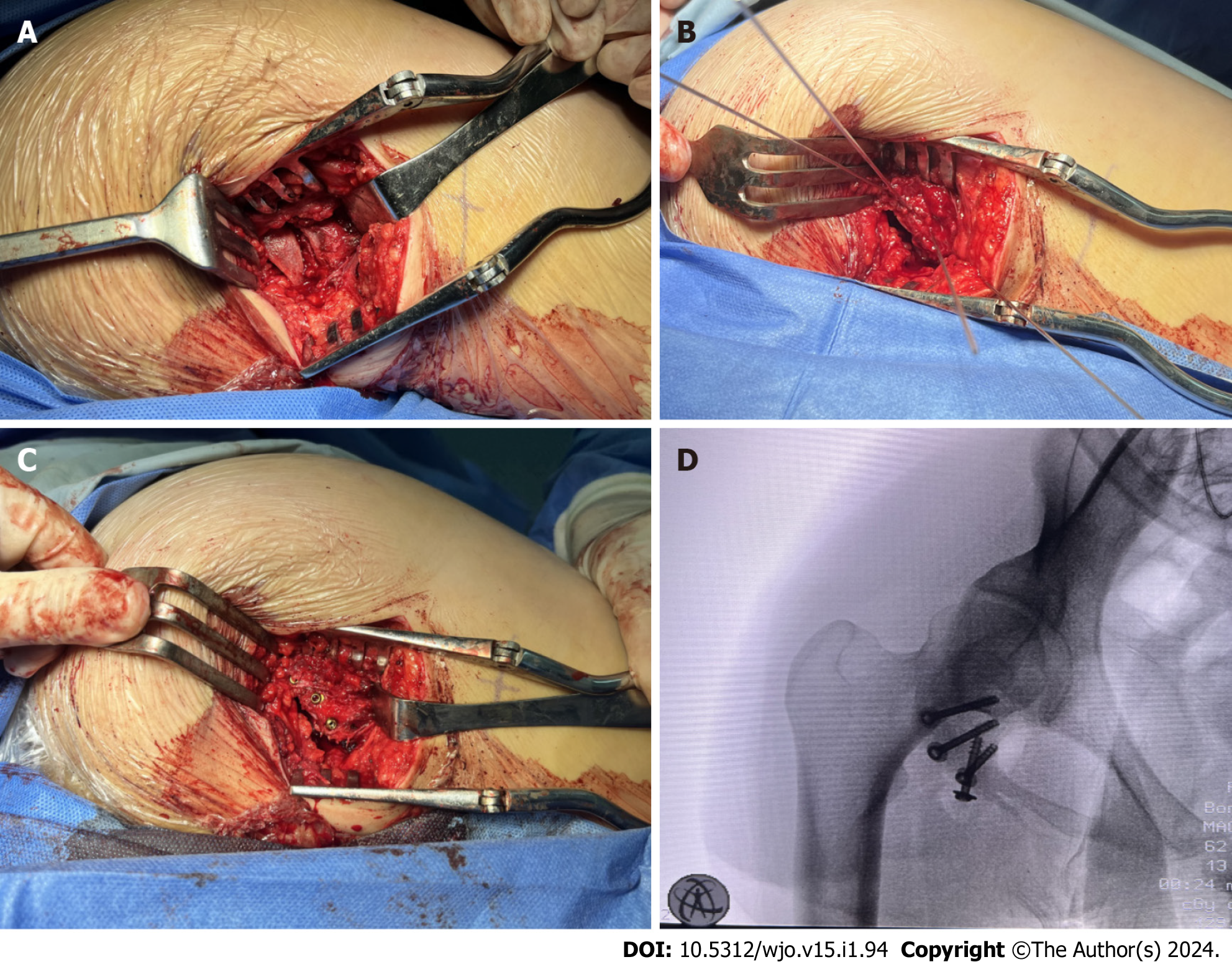
Figure 2 The photos of treatment.
A: The fracture was exposed and the fragment was seen anterolaterally displaced during the surgery; B: The fracture was temporarily fixed with Kirschner wires during the surgery; C: Four φ 4.0-mm cannulated compression screws were inserted to fix the fracture; D: C-arm fluoroscopy displayed that the fracture was properly reduced and the screws were an appropriate length and in appropriate position.
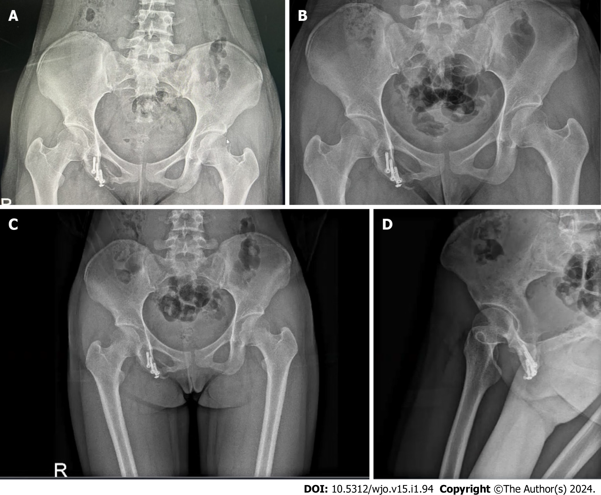
Figure 3 The results of outcome and follow-up.
A: The radiograph revealed the fracture line was fuzzy, but heterotopic ossification occurred surrounding the fracture at the 3-mo follow-up after surgery; B: The X-ray film at the 6-mo follow-up after surgery revealed that the fracture healed well without exacerbation of heterotopic ossification; C and D: The X-ray film at the 16-mo follow-up after surgery revealed that the fracture had fully healed.
- Citation: Chen ZR, Liao SJ, Yang FC. Surgical treatment of an old avulsion fracture of the ischial tuberosity and ischial ramus: A case report. World J Orthop 2024; 15(1): 94-100
- URL: https://www.wjgnet.com/2218-5836/full/v15/i1/94.htm
- DOI: https://dx.doi.org/10.5312/wjo.v15.i1.94


