Copyright
©The Author(s) 2023.
World J Orthop. Apr 18, 2023; 14(4): 207-217
Published online Apr 18, 2023. doi: 10.5312/wjo.v14.i4.207
Published online Apr 18, 2023. doi: 10.5312/wjo.v14.i4.207
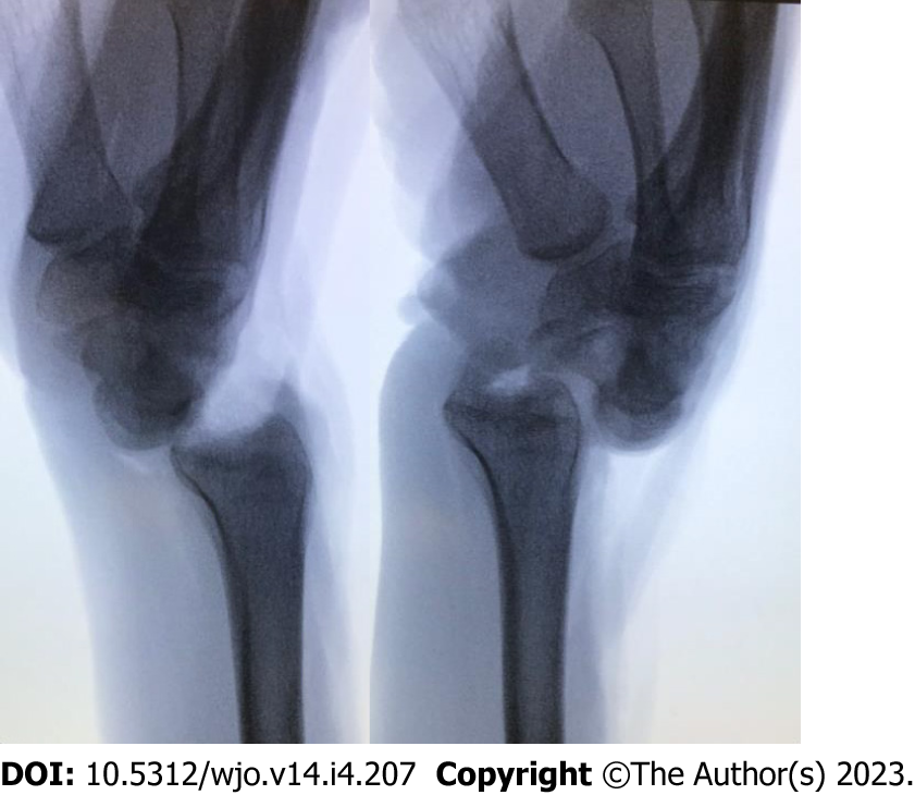
Figure 1 Lateral fluoroscopic view demonstrating complete radiocarpal instability following transection of dorsal and volar radiocarpal ligaments.
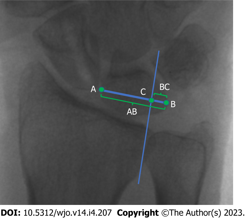
Figure 2 Gilula’s technique for measuring ulnar translocation through lunate uncovering.
A line is drawn along the transverse axis of the lunate, from the farthest radial point at the mid-portion of the lunate (point A) to its ulnar-most corner (point B). The long axis of the radius is found and a parallel line at the ulnar corner of the radius is drawn until the A-B line is transected (point C). The distance between B and C is divided by the distance between A and B (G = BC/AC)[23].
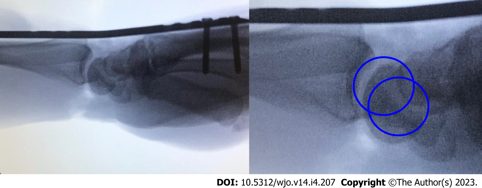
Figure 3 Concentric circle technique for assessing radiolunate joint alignment on the lateral view.
Subluxation or inadequate reduction is demonstrated by the lack of concentricity.
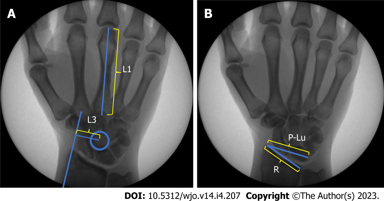
Figure 4 Chamay and Bouman techniques for measuring ulnar translocation.
A: Chamay’s index is calculated by dividing the distance between a line parallel to the axis of the radius passing through the radial styloid process and the center of rotation of the capitate (L3) and the length of the long finger metacarpal (L1)[25]; B: Bouman’s index is calculated by dividing the length of the distal articular surface of the radius (R) by the distance between the radius styloid process and the proximal ulnar corner of the lunate (P-Lu)[25].
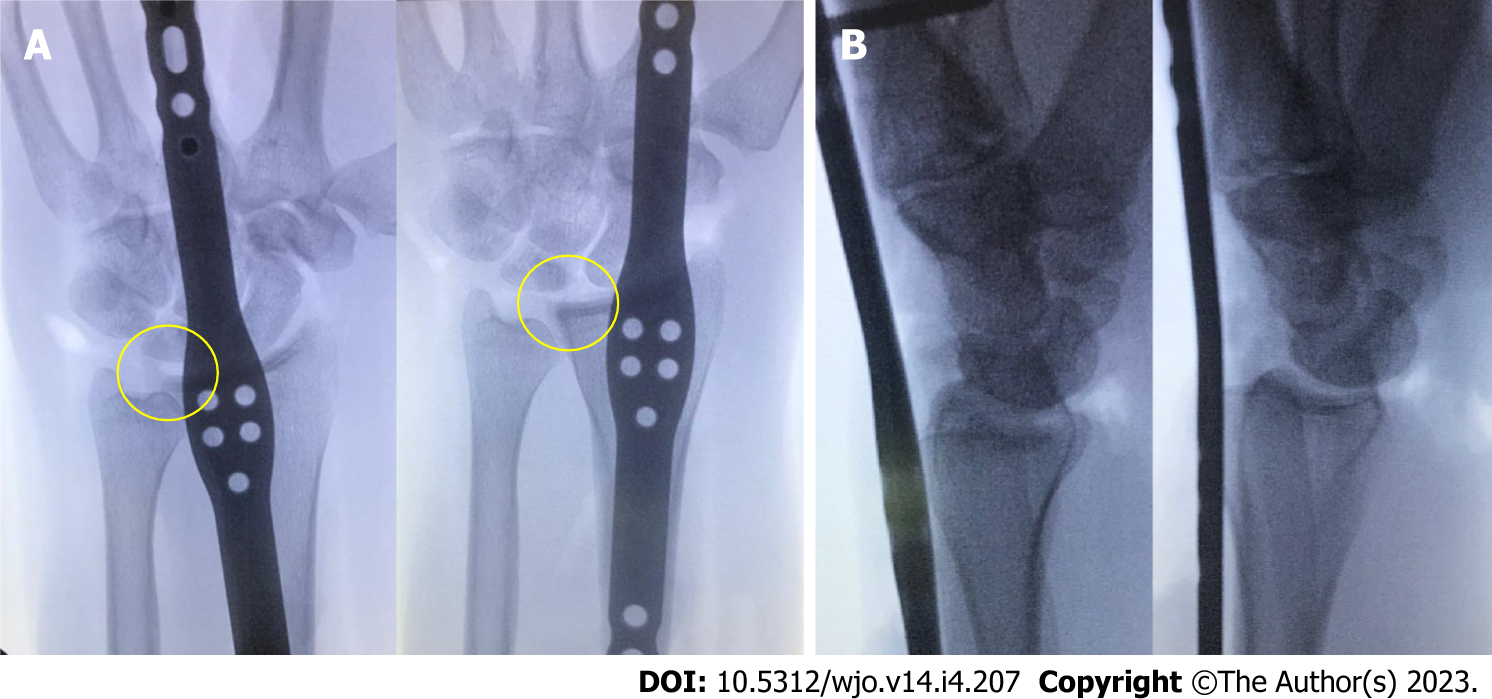
Figure 5 The effect of focusing on distal fixation alone with proximal bridge plate placement through the fourth dorsal compartment.
A: Posterior-anterior view of third metacarpal fixation (left) and second metacarpal fixation (right) demonstrates ulnar translocation of the carpus (even radial translation within the fourth dorsal compartment is insufficient to align the radiocarpal joint); B: Lateral view demonstrates volar subluxation of the radiocarpal joint, which was observed in half of the pilot study specimens with second metacarpal distal fixation (third metacarpal left, second metacarpal right).
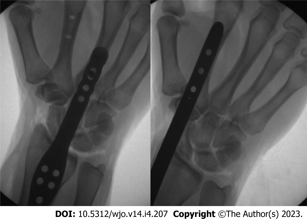
Figure 6 Anatomic radiocarpal alignment was achieved with each the second metacarpal fixation technique (2M) and third metacarpal fixation technique (3M).
- Citation: Tabeayo E, Saucedo JM, Srinivasan RC, Shah AR, Karamanos E, Rockwood J, Rodriguez-Merchan EC. Bridge plating in the setting of radiocarpal instability: Does distal fixation to the second or third metacarpal matter? A cadaveric study. World J Orthop 2023; 14(4): 207-217
- URL: https://www.wjgnet.com/2218-5836/full/v14/i4/207.htm
- DOI: https://dx.doi.org/10.5312/wjo.v14.i4.207


