Copyright
©The Author(s) 2023.
World J Orthop. Mar 18, 2023; 14(3): 113-122
Published online Mar 18, 2023. doi: 10.5312/wjo.v14.i3.113
Published online Mar 18, 2023. doi: 10.5312/wjo.v14.i3.113
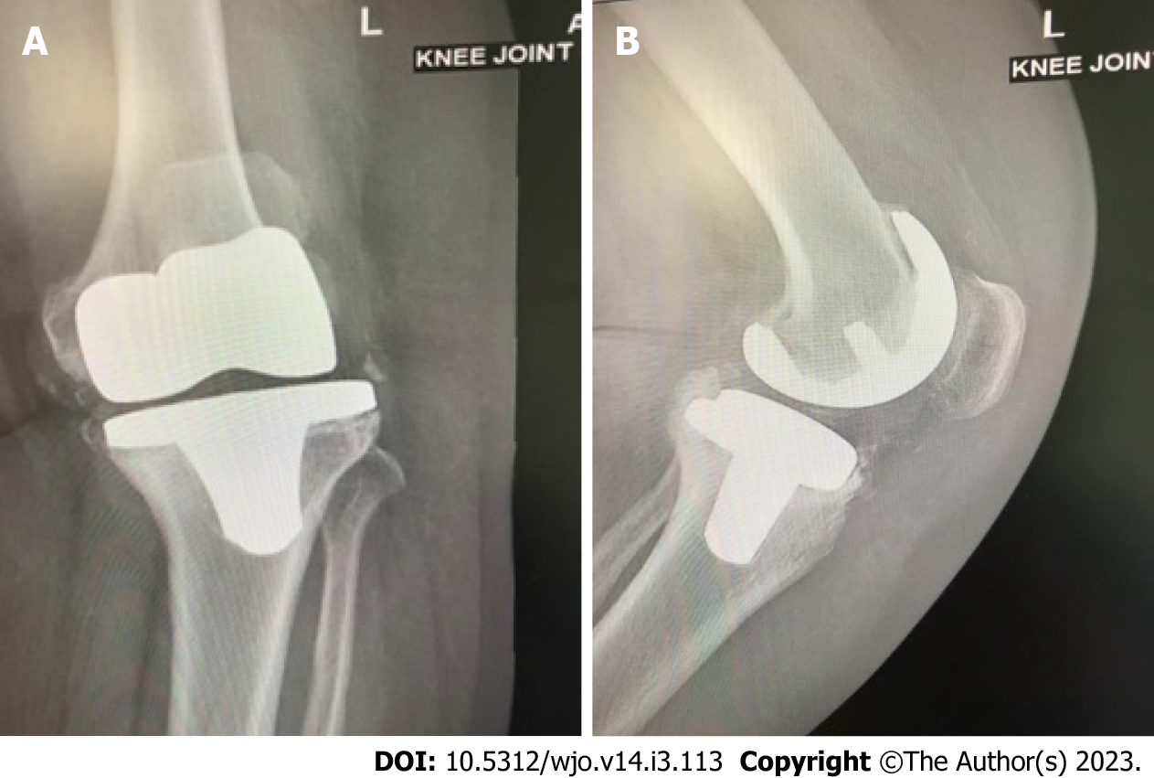
Figure 1 Anteroposterior and lateral plain radiographs showing septic loosening of a left total knee arthroplasty in a 65-year-old female patient who underwent the primary procedure 5 years previously.
A: Anteroposterior view; B: Lateral view. Preoperative aspiration confirmed Staphylococcus aureus infection and serum and synovial fluid inflammatory markers were elevated.
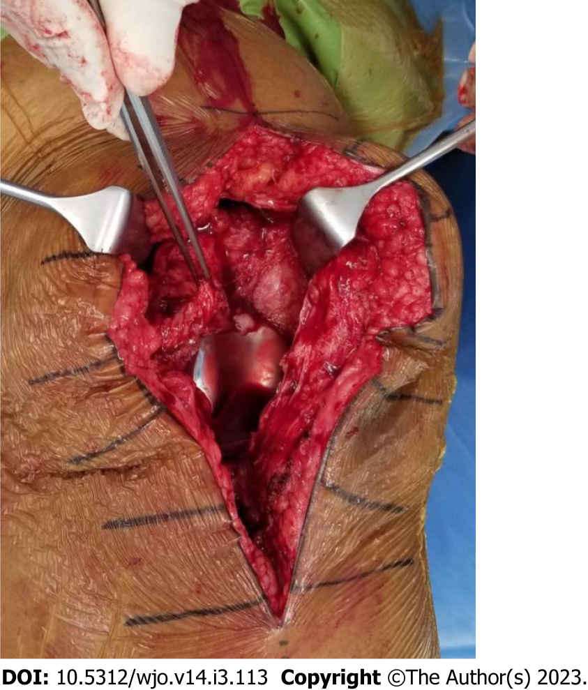
Figure 2 First stage revision of the case shown in Figure 1.
Synovial tissue surrounds the prosthesis. Total synovectomy was performed and samples sent for culture confirmed the growth of Staphylococcus aureus.
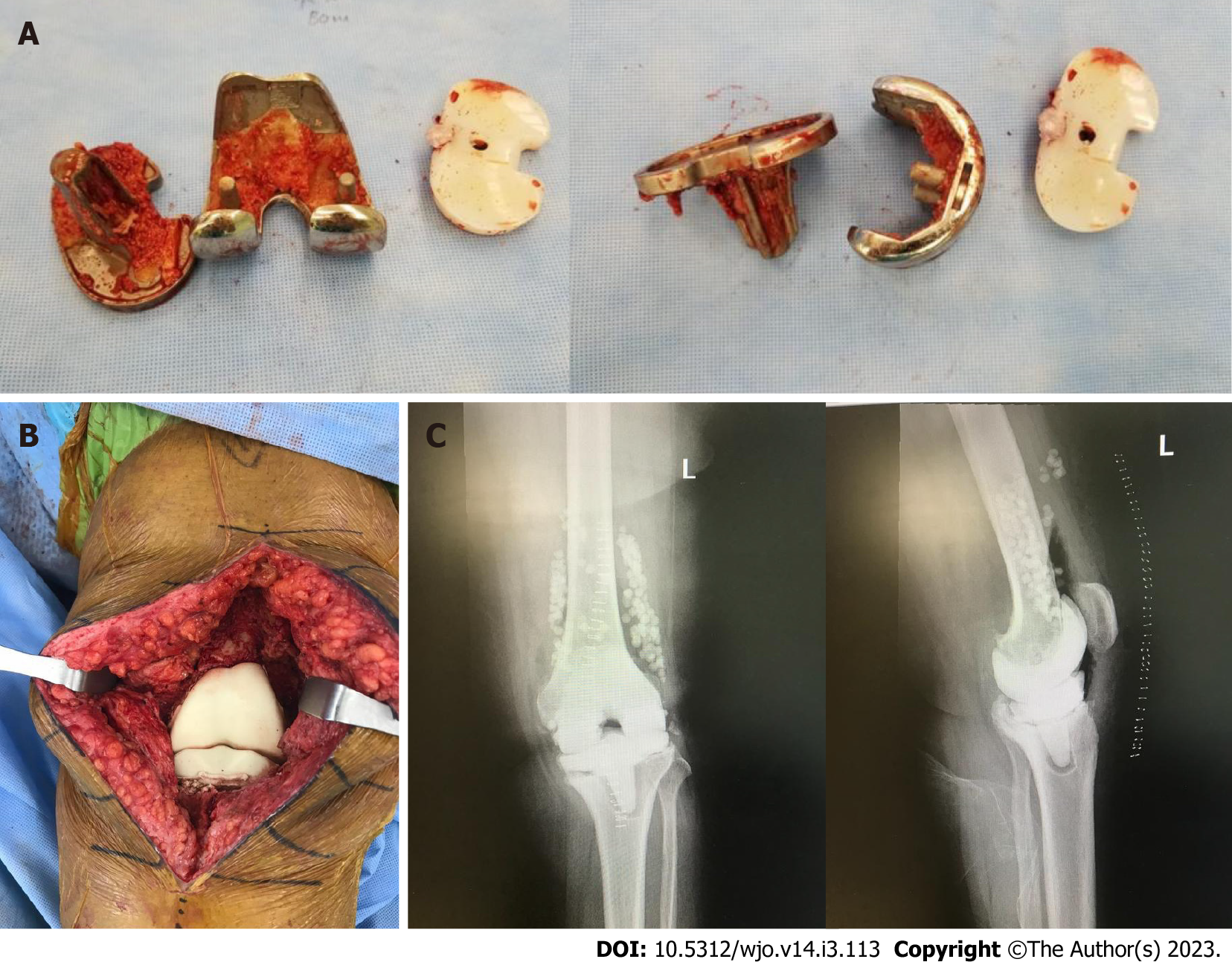
Figure 3 First stage revision.
A: Explanted prosthesis with minimal bone loss; B: Articulating spacer implantation at the end of the procedure containing vancomycin and tobramycin; C: Anteroposterior and lateral plain radiographs of the left knee after the first stage showing the spacer in situ with antibiotic beads.
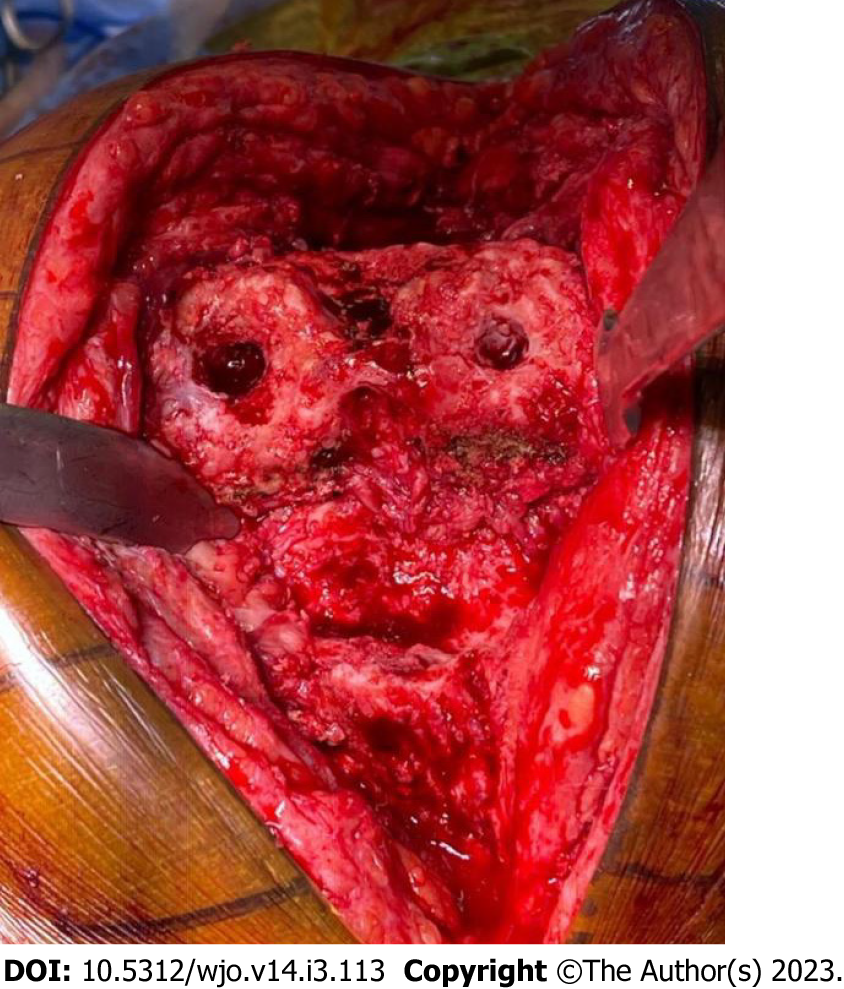
Figure 4 Second stage revision.
After the removal of the spacer, the joint is prepared for reimplantation of the prosthesis. This joint shows minimal bone loss.
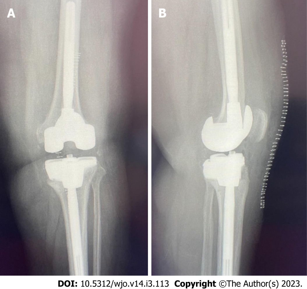
Figure 5 Postoperative anteroposterior and lateral plain radiographs.
A: Postoperative anteroposterior view of the left knee after completion of the second stage revision; B: Lateral view of the left knee after completion of the second stage revision.
- Citation: Alrayes MM, Sukeik M. Two-stage revision in periprosthetic knee joint infections. World J Orthop 2023; 14(3): 113-122
- URL: https://www.wjgnet.com/2218-5836/full/v14/i3/113.htm
- DOI: https://dx.doi.org/10.5312/wjo.v14.i3.113


