Copyright
©The Author(s) 2023.
World J Orthop. Mar 18, 2023; 14(3): 103-112
Published online Mar 18, 2023. doi: 10.5312/wjo.v14.i3.103
Published online Mar 18, 2023. doi: 10.5312/wjo.v14.i3.103
Figure 1 Image of the radiocarpal joint.
A: The crystals seen are destructing the lunate; B: Debridement of a central triangular fibro cartilage complex tear; C: The pannus seen are destructing the cartilage of distal radius; D: Ganglion stalk seen protruding into the joint; E: Resection of the ganglion stalk; F: Arthrolysis was performed by shaving the fibrous tissue inside the radiocarpal joint.
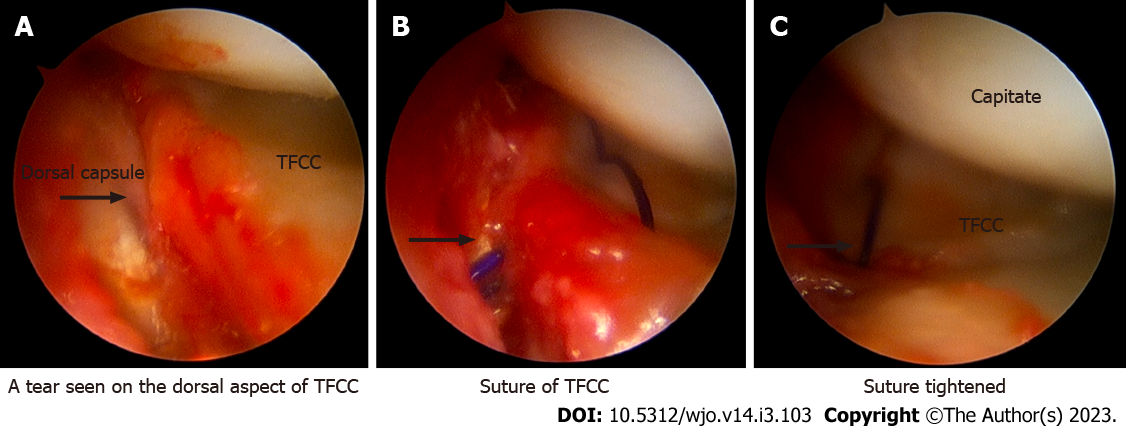
Figure 2 Repair of triangular fibro cartilage complex tear.
A: Tear seen on the dorsal aspect of the triangular fibro cartilage complex (TFCC); B: Suture of TFCC; C: Suture tightened.
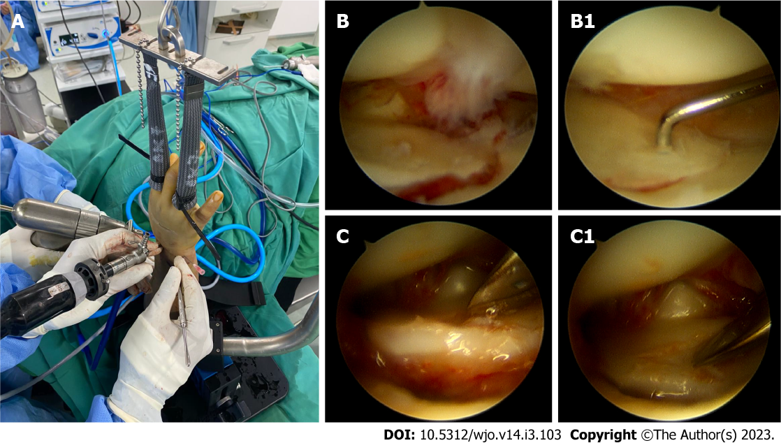
Figure 3 Arthroscopic-assisted intra-articular reduction of a distal radius fracture.
A: Finger traction was applied to the wrist; B and C: Image of intraarticular step before reduction, B1 and C1: After intraarticular reduction.
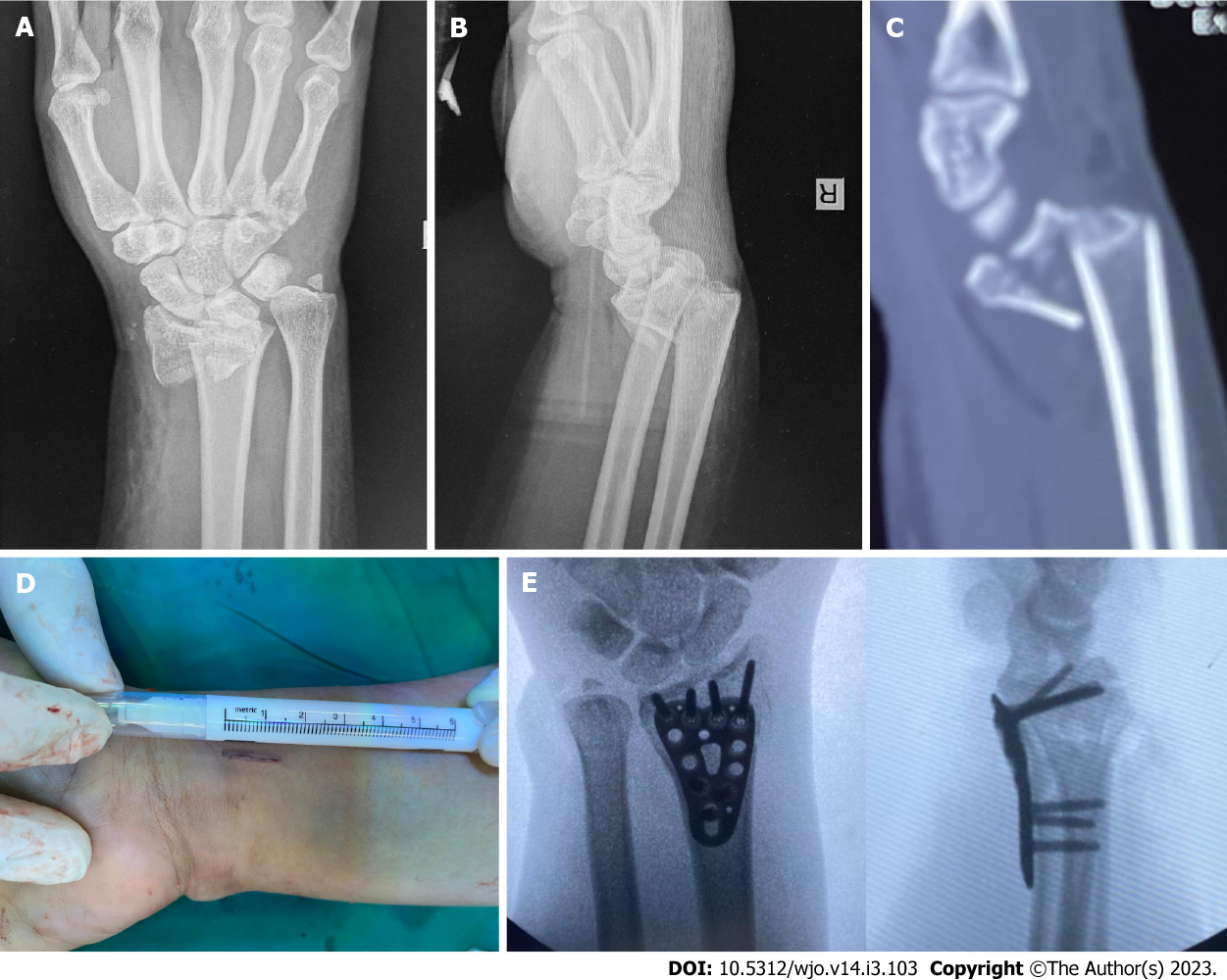
Figure 4 Preoperative X-ray and computed tomography scan showed intra-articular fragment of a distal radius fracture and postoperative X-ray of the minimally invasive plate osteosynthesis technique with a 15 mm incision.
A: Postero-anterior plain X-ray of the right wrist; B: Lateral plain X-ray of the right wrist; C: Sagittal view of computed tomography scan showed displaced intra-articular fracture of the right wrist; D: A 15 mm incision was used for the procedure; E: Postero-anterior and lateral views showed the result after plate and screw fixation.
Figure 5 Intraoperative technique of arthroscopic treatment of scaphoid nonunion.
A: Arthroscopic procedure in scaphoid nonunion graft and fixation; B: Headless screw insertion through volar percutaneous approach; C and D: Minimal wound from the arthroscopic procedure.
Figure 6 Arthroscopic view of nonunion scaphoid treatment.
A: The nonunion site was debrided; B: Debridement continued until punctate bleeding was observed; C: Nonunion site was clearly visible; D: The nonunion site was packed with bone graft.
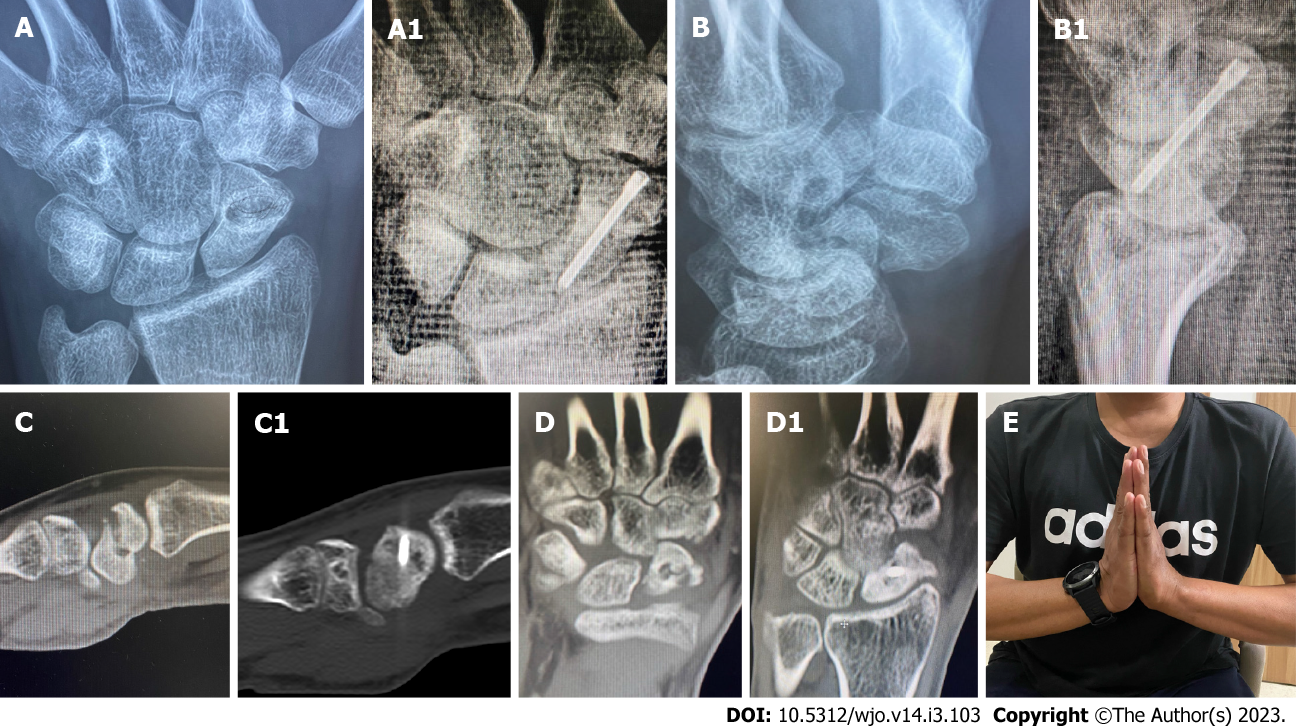
Figure 7 Arthroscopic fixation and bone grafting in scaphoid non-union.
A and B: Pre-operative X-rays; C and D: Computed tomography scans of nonunion scaphoid, showing no bony bridge and humpback deformity. The nonunion and humpback deformity was successfully healed and corrected (A1, B1, C1 and D1); E: Post-operative clinical image after five months demonstrating good and painless range of motion.
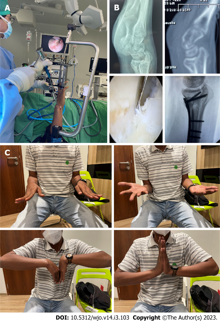
Figure 8 Arthroscopic assisted osteotomy of distal radius intra-articular malunion.
A: Arthroscopic-assisted intra-articular osteotomy; B: Pre-operative X-ray, showing volar shear intra-articular malunion with arthroscopic-assisted intra-articular osteotomy through the fracture line, and post-operative X-ray showing anatomical reduction; C: Post-operative clinical image after five months showing good and painless range of motion.
- Citation: Satria O, Hadinoto SA, Fathurrahman I. Advances in wrist arthroscopic surgery in Indonesia. World J Orthop 2023; 14(3): 103-112
- URL: https://www.wjgnet.com/2218-5836/full/v14/i3/103.htm
- DOI: https://dx.doi.org/10.5312/wjo.v14.i3.103


