Copyright
©The Author(s) 2023.
World J Orthop. Dec 18, 2023; 14(12): 889-896
Published online Dec 18, 2023. doi: 10.5312/wjo.v14.i12.889
Published online Dec 18, 2023. doi: 10.5312/wjo.v14.i12.889
Figure 1 Patient’s preoperative evaluation.
A: Clinics; B: Radiographs; C: Liner dislocation is indicated with white arrow.
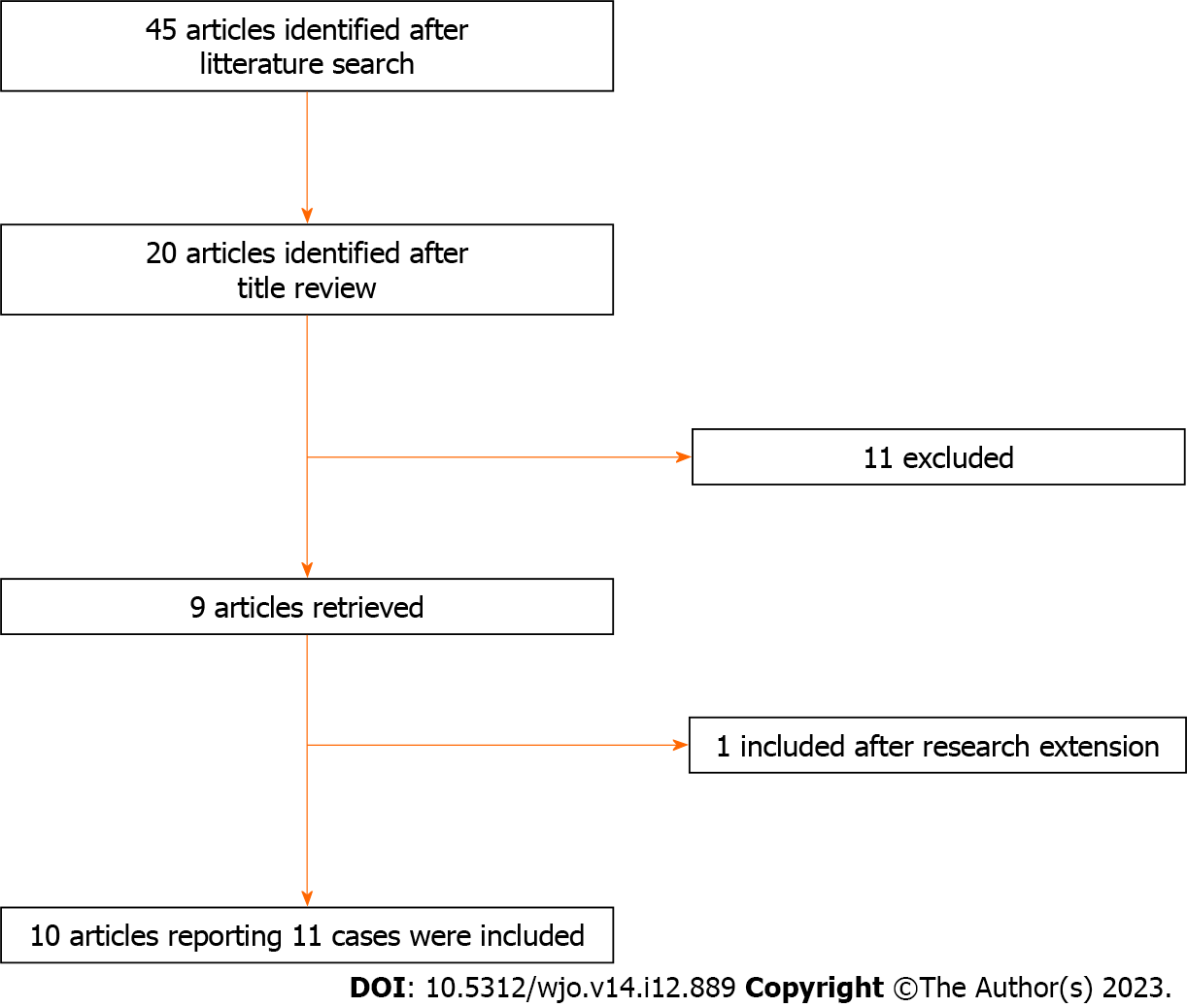
Figure 2 Summary of article inclusion process.
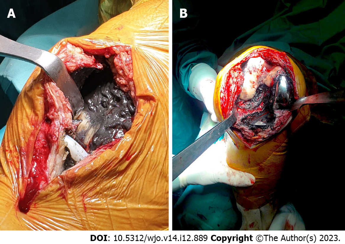
Figure 3 Intraoperative photographs documenting peri-prosthetic soft tissue metallosis.
A: Note the luxated bearing; B: Note the metal back debris.
Figure 4 Postoperative history.
A and B: Postoperative X-rays; C-E: Clinical evaluation documenting range of motion at final follow-up; F and G: Radiographies documenting implant alignment at final follow-up.
- Citation: Toro G, Braile A, Conza G, De Cicco A, Abu Mukh A, Placella G, Salini V. Unicompartimental knee arthroplasty metallosis treated with uni-on-uni revision: A case report. World J Orthop 2023; 14(12): 889-896
- URL: https://www.wjgnet.com/2218-5836/full/v14/i12/889.htm
- DOI: https://dx.doi.org/10.5312/wjo.v14.i12.889


