Copyright
©The Author(s) 2023.
World J Orthop. Oct 18, 2023; 14(10): 776-783
Published online Oct 18, 2023. doi: 10.5312/wjo.v14.i10.776
Published online Oct 18, 2023. doi: 10.5312/wjo.v14.i10.776
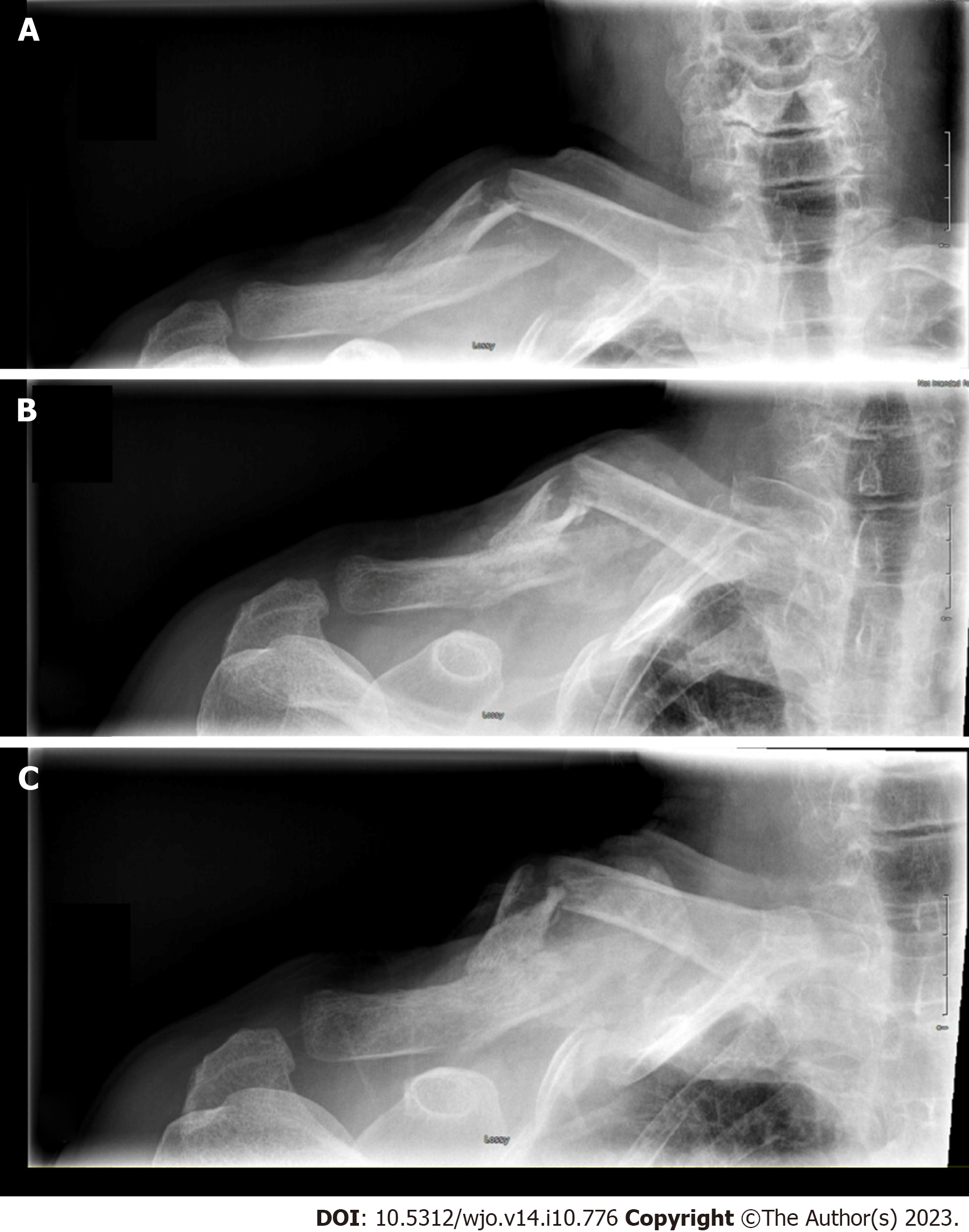
Figure 1 Antero-posterior right clavicle radiograph 2 wk, 6 wk and 3 mo post-injury.
A: 2 wk; B: 6 wk; C: 3 mo.
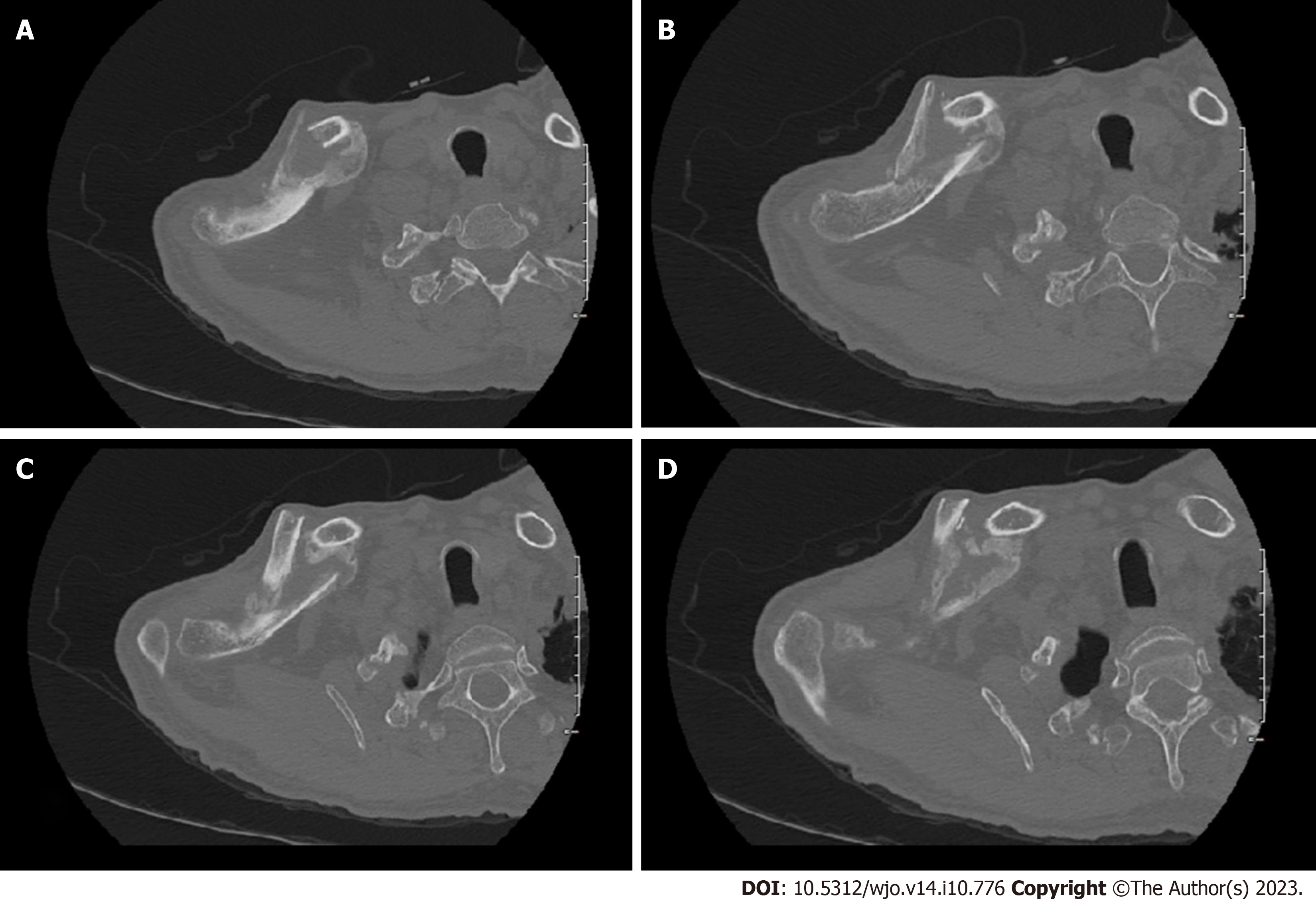
Figure 2 Sequential axial images of non-contrast computed tomography scan.
A-D: At 3 mo post-injury showing mid-shaft clavicle fracture with abundant callus formation.
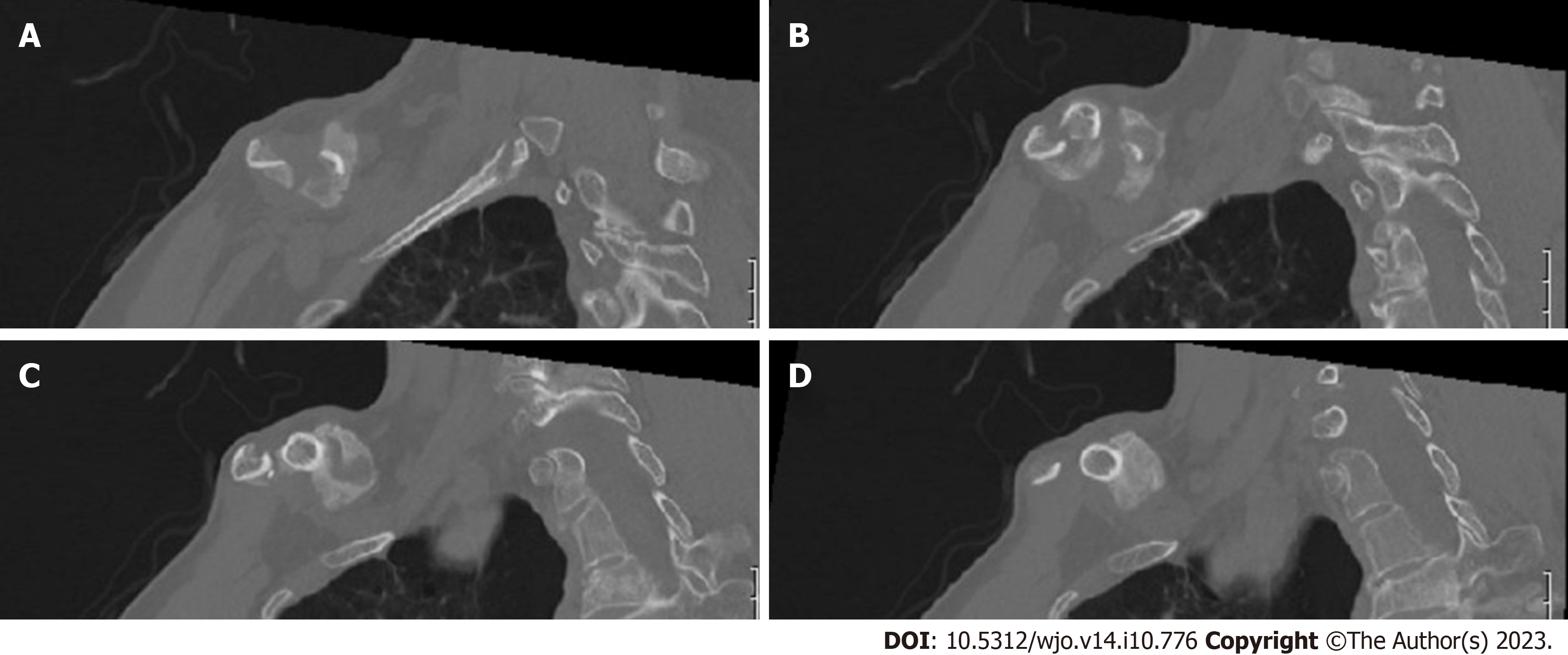
Figure 3 Sequential sagittal images of non-contrast computed tomography scan.
A-D: At 3 mo post-injury showing mid-shaft clavicle fracture with abundant callus formation.
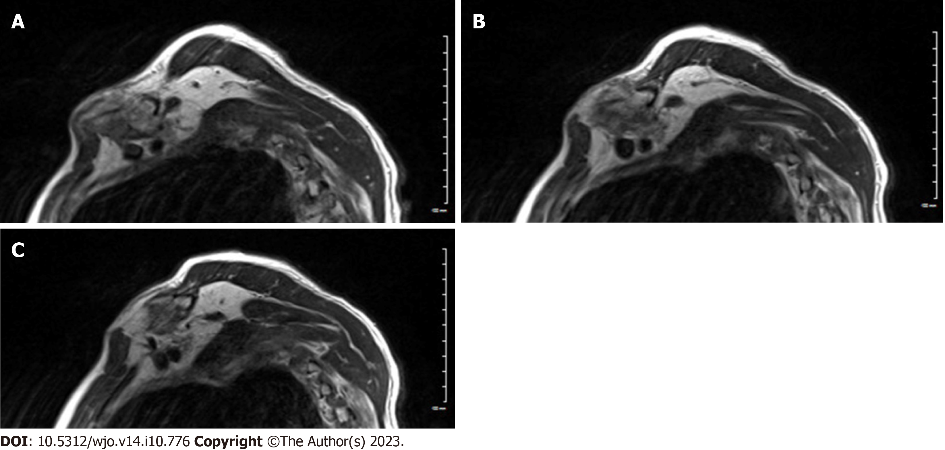
Figure 4 Sequential sagittal T2 weighted images of magnetic resonance imaging scan.
A-C: At 3 mo post-injury showing mid-shaft clavicle fracture with abundant callus formation causing a mass effect on adjacent brachial plexus trunks.
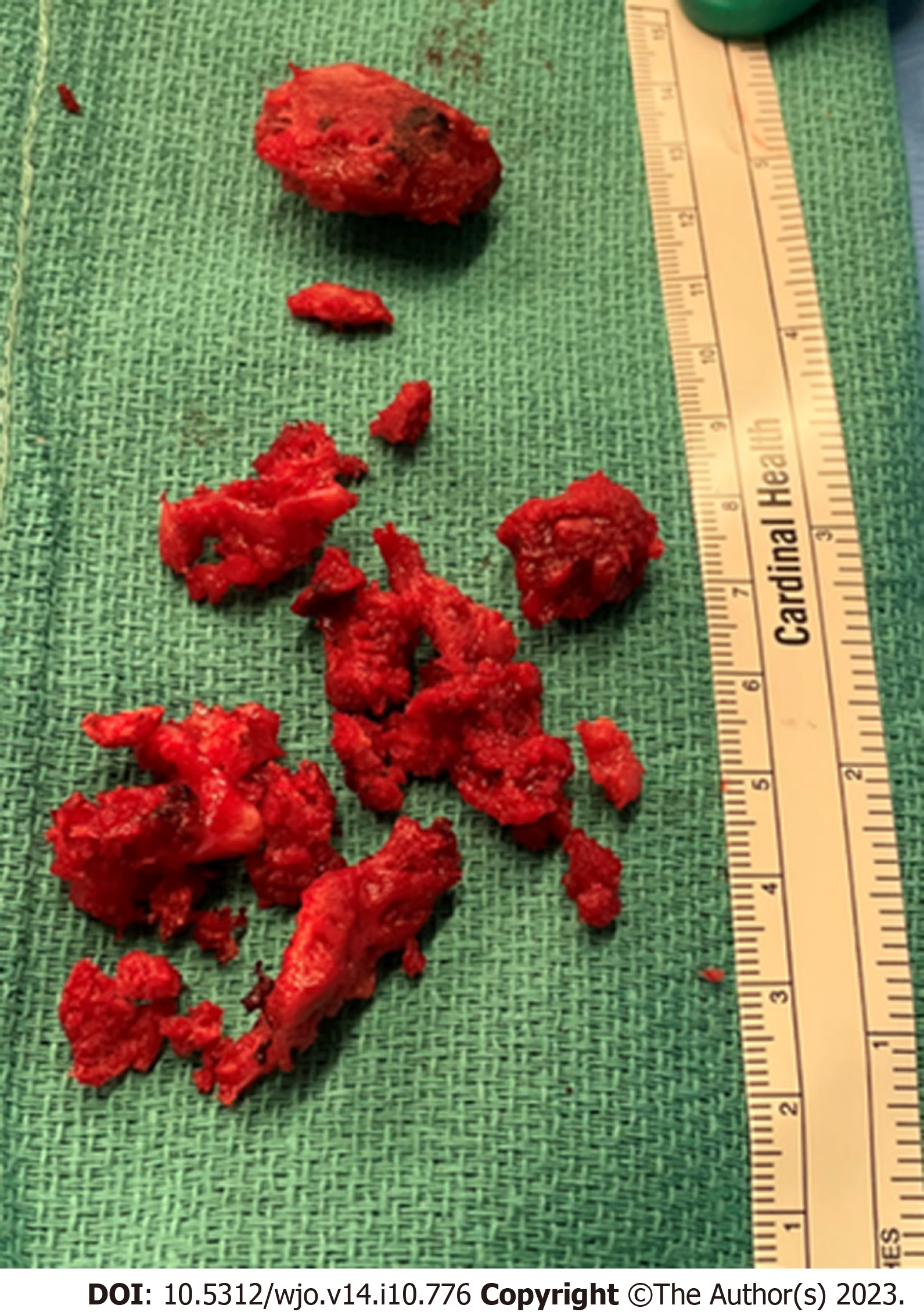
Figure 5 Clinical intra-operative picture showing the excised hypertrophied callus after surgical decompression.
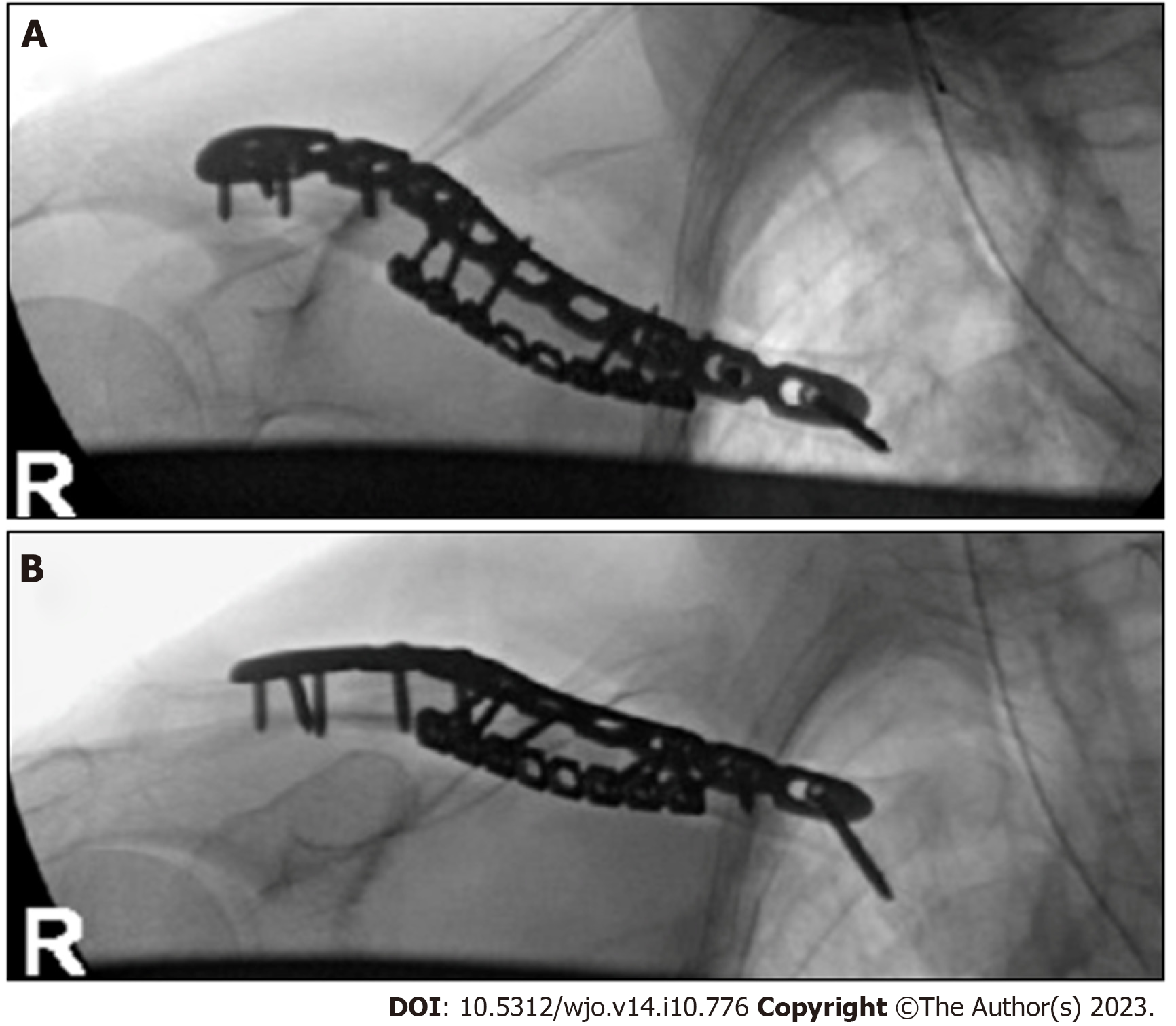
Figure 6 Superior and anteroposterior radiographs of right clavicle after open reduction and internal fixation.
A: Superior radiographs; B: Anteroposterior radiographs.
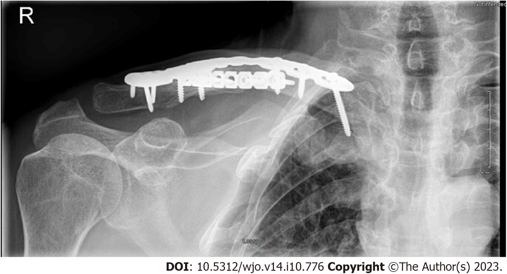
Figure 7 Anteroposterior radiographs of right clavicle at 3 mo post-surgery showing progression of the fracture healing and maintained implant fixation.
- Citation: Alzahrani MM. Late brachial plexopathy after a mid-shaft clavicle fracture: A case report. World J Orthop 2023; 14(10): 776-783
- URL: https://www.wjgnet.com/2218-5836/full/v14/i10/776.htm
- DOI: https://dx.doi.org/10.5312/wjo.v14.i10.776


